The lymphatic system, a vital component of the body’s immune and circulatory framework, helps maintain fluid balance, transports lymph, and defends against pathogens through a network of vessels and nodes. This article explores a detailed image of the lymphatic system, highlighting its key anatomical features and their roles in supporting overall health and immunity.
Lymph Lymph is a clear fluid derived from interstitial fluid, carrying white blood cells and waste products through lymphatic vessels. It plays a crucial role in returning excess fluid to the bloodstream and supporting immune responses.
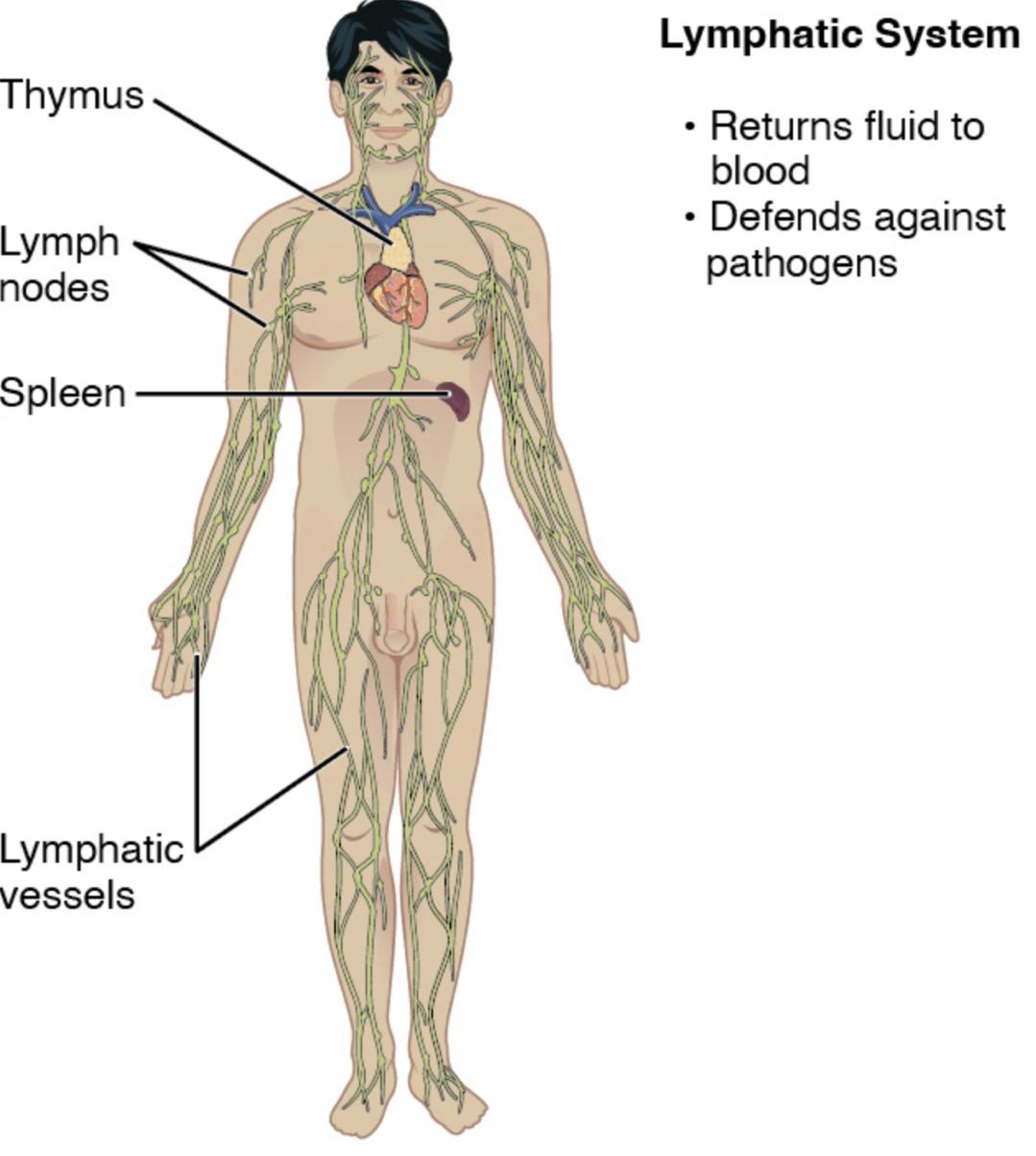
Lymphatic vessels Lymphatic vessels form a network that transports lymph from tissues back to the bloodstream, equipped with valves to prevent backflow. These vessels collect lymph from various body regions and converge toward larger ducts.
Lymph nodes Lymph nodes are small, bean-shaped structures along lymphatic vessels that filter lymph and house immune cells. They trap pathogens and initiate immune responses, swelling during infection.
Spleen The spleen, located in the upper left abdomen, filters blood, removes old red blood cells, and stores immune cells. It also produces lymphocytes to enhance immune defense.
Thymus The thymus, found in the upper chest, is crucial for maturing T-lymphocytes during early development. It shrinks with age but remains important for immune system establishment.
Tonsils Tonsils are lymphoid tissues in the throat that protect against ingested or inhaled pathogens. They form part of the body’s first line of immune defense in the oral and nasal cavities.
Peyer’s patches Peyer’s patches are lymphoid nodules in the small intestine that monitor and respond to intestinal pathogens. They play a key role in gut-associated lymphoid tissue (GALT) immunity.
Bone marrow Bone marrow produces lymphocytes and other blood cells, serving as the primary site for immune cell generation. It supports the lymphatic system by supplying new immune cells.
Thoracic duct The thoracic duct is the largest lymphatic vessel, draining lymph from the lower body and left side into the bloodstream via the left subclavian vein. It collects lymph from most of the body.
Right lymphatic duct The right lymphatic duct drains lymph from the right upper body and right side of the head into the right subclavian vein. It is smaller than the thoracic duct but equally vital.
Anatomical Overview of the Lymphatic System
The lymphatic system complements the circulatory system with a unique network of vessels and organs. This structure ensures fluid balance and immune surveillance.
- Lymph originates from interstitial fluid, collected by lymphatic vessels.
- Lymph nodes filter lymph, housing immune cells to combat infections.
- The spleen and thymus contribute to blood filtration and immune cell maturation.
- Tonsils and Peyer’s patches provide localized immune protection.
- Bone marrow serves as the source of new lymphocytes.
Role of Lymphatic Vessels and Ducts
Lymphatic vessels and ducts form the transport system for lymph. This network maintains fluid dynamics and immune function.
- Lymphatic vessels collect excess fluid from tissues, preventing edema.
- Valves in these vessels ensure unidirectional lymph flow.
- The thoracic duct drains the majority of the body’s lymph.
- The right lymphatic duct handles the right upper quadrant drainage.
- These ducts empty into the subclavian veins, returning lymph to circulation.
Functions of Lymphoid Organs
Lymphoid organs play diverse roles in immunity and blood filtration. Their strategic locations enhance protective capabilities.
- The spleen filters blood, removing damaged cells and pathogens.
- The thymus matures T-cells, critical for adaptive immunity.
- Tonsils guard the throat against respiratory pathogens.
- Peyer’s patches monitor the gut for ingested threats.
- Bone marrow produces B-cells and T-cells for immune responses.
Clinical Relevance and Associated Conditions
Understanding lymphatic anatomy aids in diagnosing and treating related disorders. This knowledge supports effective medical interventions.
- Blockage of lymphatic vessels can lead to lymphedema, causing swelling.
- Enlarged lymph nodes may indicate lymphoma or infection.
- Spleen enlargement, or splenomegaly, can signal underlying disease.
- Thymus abnormalities are linked to immune deficiency syndromes.
- Imaging and biopsies assess lymphatic system health.
The lymphatic system, with its intricate network of lymph, lymphatic vessels, nodes, and organs like the spleen, thymus, tonsils, Peyer’s patches, and bone marrow, is essential for fluid balance and immune defense. The thoracic and right lymphatic ducts ensure efficient drainage, while conditions like lymphedema highlight the importance of this system’s integrity. This detailed exploration provides a solid foundation for understanding lymphatic anatomy and addressing related health challenges.

