The forearm is a critical region of the upper limb, housing deep muscles that play a pivotal role in the intricate movements of the wrists, hands, and fingers. This article delves into the anatomy of the left forearm deep muscles as depicted in a palmar view, offering a detailed examination of their structure, origins, and functions. The provided image serves as an essential tool for understanding the deeper muscular layers that support fine motor skills and inform clinical interventions.
Introduction to the Image
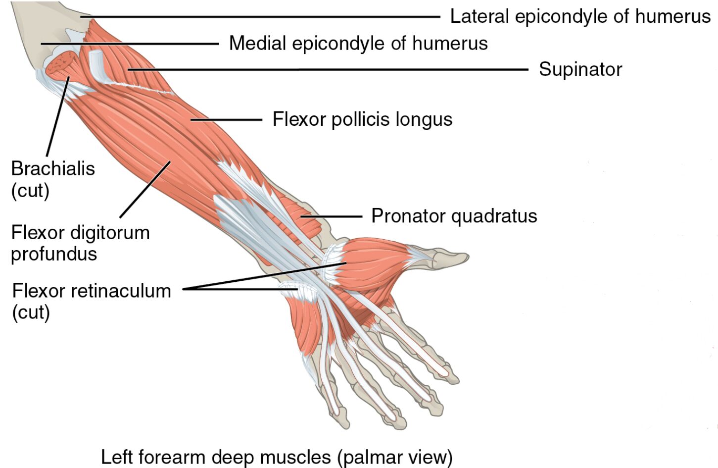
Investigating the deeper layers of the forearm reveals its complex design. The image illustrates the left forearm deep muscles in a palmar view, with clear labels identifying each component.
- Medial epicondyle of humerus: A bony prominence on the humerus, serving as an attachment site for flexor muscles.
- Lateral epicondyle of humerus: Another humeral landmark, providing anchorage for extensor muscles.
- Supinator: Originating from the lateral epicondyle and ulna, it supinates the forearm by rotating the radius.
- Flexor pollicis longus: Arising from the radius, it flexes the thumb at the interphalangeal joint.
- Brachialis (cut): Originating from the humerus, it flexes the elbow and is partially sectioned for visibility.
- Flexor digitorum profundus: Stemming from the ulna and interosseous membrane, it flexes the distal phalanges of the fingers.
- Pronator quadratus: Arising from the ulna, it pronates the forearm by rotating the radius.
- Flexor retinaculum (cut): A fibrous band, sectioned here, that stabilizes the flexor tendons at the wrist.
Anatomical Overview
Delving into the deep muscular structure uncovers a sophisticated network. The medial epicondyle of humerus and lateral epicondyle of humerus act as attachment points, while the supinator, flexor pollicis longus, brachialis (cut), flexor digitorum profundus, pronator quadratus, and flexor retinaculum (cut) form the deep layer, driving precise forearm and finger movements.
- The medial and lateral epicondyles of humerus provide stable anchor points for muscle origins.
- The supinator enables forearm supination, rotating the palm upward.
- The flexor pollicis longus facilitates thumb flexion, crucial for gripping.
- The brachialis (cut) supports elbow flexion, revealed by its partial sectioning.
- The flexor digitorum profundus allows flexion of the distal finger joints.
- The pronator quadratus drives forearm pronation, rotating the palm downward.
- The flexor retinaculum (cut) stabilizes tendons, preventing bowstringing during wrist movement.
Functional Roles of Deep Forearm Muscles
Understanding the functional contributions highlights their importance in movement. These muscles work in concert to execute specialized actions, from supinating the forearm to flexing the fingers, relying on their deep positioning.
- The supinator facilitates supination, essential for activities like turning a doorknob.
- The flexor pollicis longus enables thumb flexion, vital for precise grasping.
- The brachialis (cut) provides strong elbow flexion, supporting lifting tasks.
- The flexor digitorum profundus allows distal finger flexion, key for gripping objects.
- The pronator quadratus drives pronation, important for rotational movements.
- The flexor retinaculum (cut) maintains tendon alignment, enhancing wrist stability.
Clinical Significance
Examining the clinical relevance underscores their practical value. Injuries or dysfunctions in these deep muscles can impair hand and forearm function, necessitating targeted therapeutic approaches.
- Strain in the supinator can limit supination, often managed with physical therapy.
- The flexor pollicis longus injury may reduce thumb strength, requiring rehabilitation.
- The brachialis (cut) damage can weaken elbow flexion, treated with rest and exercises.
- The flexor digitorum profundus dysfunction can impair finger flexion, affecting dexterity.
- The pronator quadratus strain may cause pronation weakness, needing strengthening routines.
- The flexor retinaculum (cut) issues, such as carpal tunnel syndrome, can compress nerves, requiring surgical intervention.
Conclusion
The exploration of left forearm deep muscles in a palmar view reveals a remarkable interplay of anatomy and function. The medial epicondyle of humerus, lateral epicondyle of humerus, supinator, flexor pollicis longus, brachialis (cut), flexor digitorum profundus, pronator quadratus, and flexor retinaculum (cut) each contribute uniquely to forearm rotation, finger flexion, and wrist stability. This understanding not only enhances appreciation of the forearm’s deeper structure but also supports effective management of related conditions, improving overall limb functionality and health.

