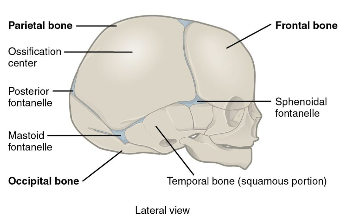
The lateral view of a newborn skull offers a detailed insight into the unique cranial anatomy of infants, characterized by soft spots and developing bones. This structure allows for flexibility during birth and accommodates rapid brain growth in the early stages of life, making it a critical area for understanding pediatric anatomy.
Labels Introduction
Parietal bone: The parietal bone forms the majority of the skull’s sides and roof, consisting of two large, curved plates that meet at the sagittal suture. These bones protect the brain and allow for slight movement during birth due to their incomplete fusion.
Ossification center: The ossification center is the initial site where bone formation begins within the cartilage model of the skull, gradually hardening over time. This process starts prenatally and continues postnatally, contributing to the skull’s strength and stability.
Posterior fontanelle: The posterior fontanelle is a small, diamond-shaped soft spot at the junction of the parietal and occipital bones, closing within a few months after birth. It allows the skull to compress during delivery and permits brain expansion in the early developmental phase.
Mastoid fontanelle: The mastoid fontanelle is a small, irregular soft spot located near the junction of the parietal, occipital, and temporal bones, typically closing by the end of the first year. It provides additional flexibility during birth and serves as a site for monitoring intracranial pressure.
Occipital bone: The occipital bone forms the back and base of the skull, housing the foramen magnum where the spinal cord exits, and protects the cerebellum. In newborns, it remains partially unfused, allowing for cranial molding during delivery.
Sphenoidal fontanelle: The sphenoidal fontanelle, or anterior lateral fontanelle, is a larger soft spot near the junction of the frontal, parietal, temporal, and sphenoid bones, closing around 6-12 months. It facilitates cranial deformation during birth and supports early brain growth.
Frontal bone: The frontal bone forms the forehead and upper part of the eye sockets, consisting of two halves in newborns that fuse into a single bone later. This bone protects the frontal lobes of the brain and allows for facial development.
Temporal bone (squamous portion): The temporal bone’s squamous portion forms the side of the skull above the ear, contributing to the temporal fossa and housing the middle ear structures. In newborns, this area is still developing, providing flexibility and room for brain expansion.
Detailed Anatomical Overview
Cranial Bones in Newborns
The newborn skull’s lateral view showcases a combination of bony and membranous structures, designed to adapt to the birthing process. The parietal bone and frontal bone are key components, offering protection while remaining flexible due to unfused sutures. This adaptability is crucial for accommodating the brain’s rapid growth in the first year.
- The occipital bone supports the brainstem and cerebellum, with its foramen magnum allowing neural communication.
- The temporal bone houses the developing auditory system, including the tympanic cavity.
- The sphenoidal fontanelle and mastoid fontanelle provide additional elasticity during delivery.
Role of Fontanelles and Ossification
Fontanelles are critical soft spots that allow the skull to compress and expand, aiding in childbirth and postnatal development. The posterior fontanelle closes early, typically by 2-3 months, while the ossification center marks the beginning of bone hardening. These features ensure the skull can grow with the brain without pressure.
- The sphenoidal fontanelle allows monitoring of intracranial pressure in cases of hydrocephalus.
- The mastoid fontanelle may be palpated to assess cerebrospinal fluid dynamics.
- The ossification center progresses from the center outward, strengthening the skull over time.
Clinical Significance and Growth
The newborn skull’s structure is vital for assessing developmental milestones and diagnosing conditions. The frontal bone’s two halves eventually fuse at the metopic suture, typically by age 2, shaping the forehead. Abnormal closure of fontanelles can indicate craniosynostosis, requiring medical attention.
- The parietal bone’s sutures allow overlapping during birth, reducing head trauma.
- The occipital bone’s flexibility supports head positioning in utero.
- The temporal bone’s squamous portion develops the zygomatic process, aiding facial structure.
Protective and Developmental Functions
The skull’s design protects the brain while allowing for growth, with fontanelles serving as pressure relief points. The jugular notch and mastoid fontanelle are palpated to evaluate neurological health, especially in premature infants. This balance of protection and plasticity is unique to the newborn period.
- The posterior fontanelle closes as the sagittal suture strengthens, stabilizing the skull.
- The sphenoidal fontanelle supports the sphenoid bone’s role in cranial base formation.
- The ossification center ensures gradual hardening, preventing premature rigidity.
The lateral view of the newborn skull highlights its remarkable adaptability and protective role. Its labeled components work together to support brain development and facilitate a safe delivery. A deep understanding of this anatomy enhances the ability to monitor growth and address potential health concerns effectively.
Conclusion
The lateral view of the newborn skull reveals a dynamic structure designed to protect the developing brain while allowing for growth and flexibility. The presence of fontanelles and ossification centers underscores its role in accommodating rapid neurological expansion and ensuring a safe birthing process. Exploring these features provides valuable insights into pediatric anatomy and its clinical implications.

