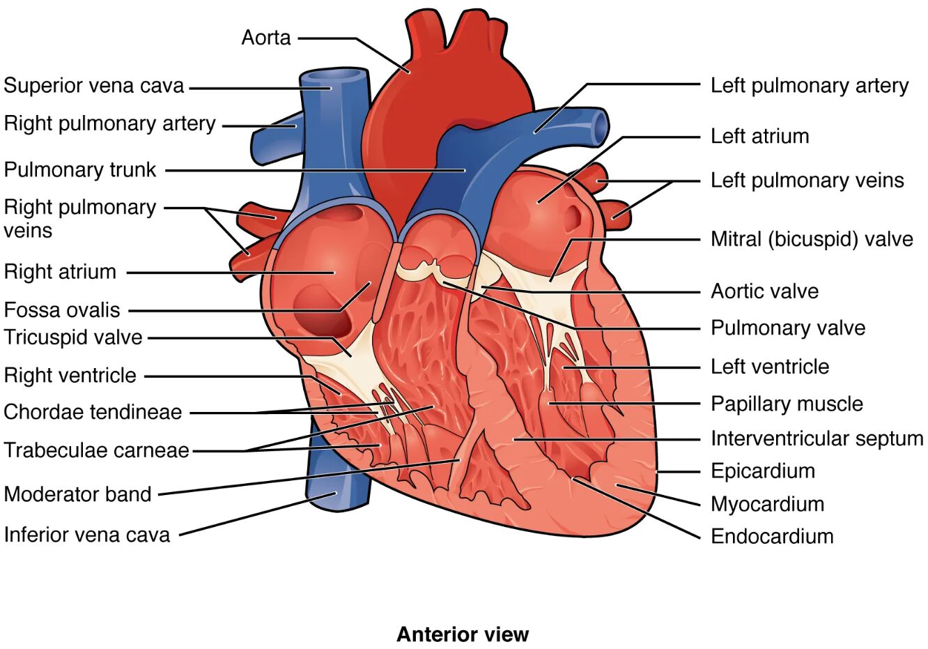
The heart’s internal anatomy is a complex network that drives its life-sustaining function, revealed vividly in this anterior view. This diagram showcases the four chambers, major vessels with their early branches, and the critical valves, with the pulmonary trunk and aorta partially obscuring the interatrial septum and the atrioventricular septum cut away for clarity. Exploring this image provides a deeper appreciation of how the heart coordinates blood flow through its intricate internal structures.
Labelled Parts Explanation
- Right atrium The right atrium receives deoxygenated blood from the superior and inferior vena cava, serving as the entry point for the right side of the heart. It contracts to push blood through the tricuspid valve into the right ventricle.
- Right ventricle The right ventricle pumps deoxygenated blood into the pulmonary trunk for oxygenation in the lungs, featuring a thinner myocardium suited to lower pressure. Its internal trabeculae carneae enhance its contractile efficiency.
- Left atrium The left atrium collects oxygenated blood from the pulmonary veins, preparing it for systemic circulation. It contracts to move blood through the mitral valve into the left ventricle.
- Left ventricle The left ventricle ejects oxygenated blood into the aorta, with its thick myocardium generating high pressure to overcome systemic resistance. Its internal structure includes papillary muscles that anchor the mitral valve.
- Pulmonary trunk The pulmonary trunk carries deoxygenated blood from the right ventricle to the lungs, dividing into the left and right pulmonary arteries. It is a critical conduit in the pulmonary circulation pathway.
- Aorta The aorta distributes oxygenated blood from the left ventricle to the systemic circulation, branching into major arteries like the coronary and brachiocephalic arteries. Its initial segment, the ascending aorta, is visible in this view.
- Tricuspid valve The tricuspid valve separates the right atrium from the right ventricle, allowing unidirectional blood flow during atrial contraction. Its three cusps close to prevent backflow into the atrium during ventricular systole.
- Mitral valve The mitral valve, or bicuspid valve, lies between the left atrium and left ventricle, ensuring blood flows toward the ventricle. Its two cusps are supported by chordae tendineae to prevent prolapse during contraction.
- Interatrial septum The interatrial septum is the wall dividing the right and left atria, maintaining separation of oxygenated and deoxygenated blood. It contains the fossa ovalis, a remnant of the fetal foramen ovale.
- Atrioventricular septum The atrioventricular septum separates the atria from the ventricles, providing structural support and housing the atrioventricular node. Its partial removal in this view exposes the atrioventricular valves.
- Papillary muscles The papillary muscles are conical projections within the ventricles, anchoring the chordae tendineae to the tricuspid and mitral valves. They prevent valve inversion during ventricular contraction.
- Chordae tendineae The chordae tendineae are strong, fibrous cords that connect the papillary muscles to the valve cusps, stabilizing the tricuspid and mitral valves. They ensure valves close properly to maintain unidirectional blood flow.
Anatomical Overview of the Heart’s Internal Structures
The internal layout of the heart is a masterpiece of design, supporting its role as a dual pump. This anterior view reveals the chambers, vessels, and valves that orchestrate blood movement.
- The right atrium and left atrium serve as receiving chambers, separated by the interatrial septum.
- The right ventricle and left ventricle act as pumping chambers, with the atrioventricular septum providing a barrier.
- The pulmonary trunk and aorta emerge as major outlets, driving pulmonary and systemic circulation.
- Valves like the tricuspid valve and mitral valve, supported by papillary muscles and chordae tendineae, ensure efficient flow.
This structure enables the heart to maintain separate circulatory pathways with precision.
Role of the Chambers in Blood Flow
The heart’s chambers are central to its pumping mechanism. Their internal anatomy supports distinct circulatory roles.
- The right atrium receives deoxygenated blood, channeling it to the right ventricle via the tricuspid valve.
- The left atrium collects oxygenated blood, passing it to the left ventricle through the mitral valve.
- The right ventricle pumps blood into the pulmonary trunk, while the left ventricle ejects it into the aorta.
- The thicker left ventricle reflects its higher pressure demands compared to the right ventricle.
This chamber design ensures balanced circulation to the lungs and body.
Function of Major Vessels and Valves
The major vessels and valves regulate blood flow within the heart. This view highlights their critical interactions.
- The pulmonary trunk transports deoxygenated blood to the lungs, guided by the pulmonary valve (not labeled but implied).
- The aorta distributes oxygenated blood, with the aortic valve preventing backflow.
- The tricuspid valve and mitral valve open during diastole, closing during systole to maintain directionality.
- The papillary muscles and chordae tendineae stabilize these valves during contraction.
This system ensures efficient and unidirectional blood movement.
Structural Significance of the Septa
The septa provide essential division within the heart. Their anatomy supports functional separation.
- The interatrial septum prevents mixing of oxygenated and deoxygenated blood in the atria.
- The atrioventricular septum isolates the atria from the ventricles, housing the conduction system.
- The cutaway view reveals the mitral valve and tricuspid valve, emphasizing septal support.
- The interatrial septum’s fossa ovalis is a key landmark in fetal circulation studies.
These structures maintain the heart’s dual circulatory integrity.
Physiological Role of Internal Support Structures
The internal support structures enhance the heart’s pumping efficiency. Their design optimizes valve function.
- The papillary muscles contract to tense the chordae tendineae, preventing valve prolapse.
- The chordae tendineae anchor the tricuspid valve and mitral valve, ensuring tight closure.
- This mechanism supports the left ventricle and right ventricle during high-pressure ejection.
- The stability provided aids in maintaining cardiac output.
These components are vital for the heart’s rhythmic performance.
Clinical Relevance of Internal Anatomy
Understanding the heart’s internal structures aids in diagnosing and treating cardiovascular conditions. These features are key clinical markers.
- Stenosis or regurgitation of the tricuspid valve or mitral valve can impair blood flow.
- Dilation of the pulmonary trunk may indicate pulmonary hypertension.
- The interatrial septum is assessed for atrial septal defects, which can cause shunting.
- Imaging techniques evaluate the left ventricle’s thickness and valve function.
This knowledge supports effective cardiac care and intervention.
Conclusion
The internal structures of the heart, as depicted in this anterior view, offer a detailed insight into the chambers, vessels, and valves that drive circulation. By exploring the right atrium, left atrium, right ventricle, left ventricle, and their associated structures like the pulmonary trunk and aorta, one gains a comprehensive understanding of the heart’s internal dynamics. This knowledge serves as a valuable resource for studying cardiovascular anatomy and addressing related health concerns, encouraging further exploration of the heart’s intricate design and physiological significance.

