The journey of human life begins with a remarkable event called implantation, where a developing embryo establishes a secure connection with the mother’s uterus. This crucial step is elegantly illustrated in the provided diagram, offering a detailed view of the cellular interactions and transformations that occur. Understanding this process is fundamental to comprehending early pregnancy and the intricate biological symphony that supports new life.
Understanding the Implantation Process
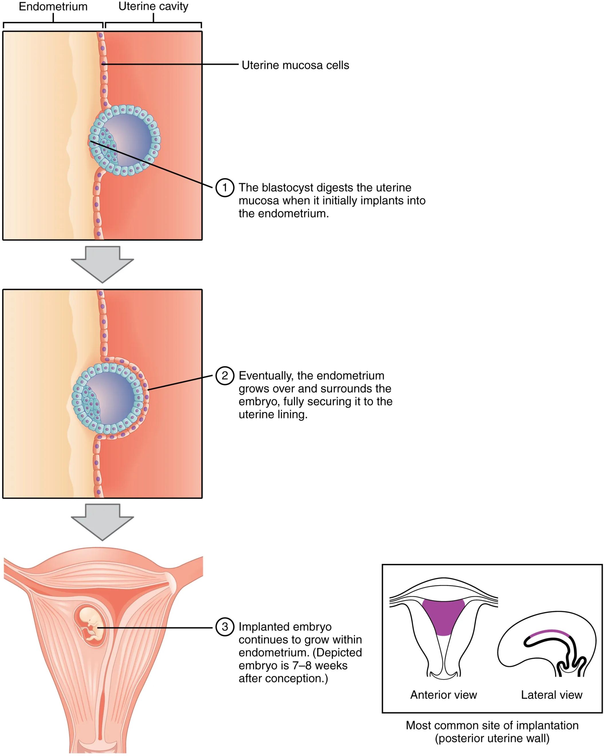
The image beautifully dissects the implantation process, highlighting the key stages from initial adhesion to full embedment within the uterine lining. This intricate dance between the blastocyst and the endometrium is essential for the successful continuation of a pregnancy.
Endometrium: This refers to the inner lining of the uterus, which plays a critical role in supporting and nourishing a developing embryo. During the menstrual cycle, the endometrium thickens in preparation for potential implantation, becoming rich in blood vessels and glandular secretions.
Uterine cavity: This is the hollow space within the uterus where the embryo implants and develops. It provides a protected environment for the growing fetus, facilitating its nourishment and waste removal throughout gestation.
Uterine mucosa cells: These are the specialized cells that form the surface layer of the endometrium, directly interacting with the invading blastocyst. Their role is pivotal in mediating the initial adhesion and subsequent embedding of the embryo.
The blastocyst digests the uterine mucosa when it initially implants into the endometrium: This annotation points to the initial stage where the outer layer of the blastocyst, the trophoblast, actively breaks down the endometrial tissue. This invasive action allows the embryo to firmly attach to the uterine lining, marking the beginning of a secure connection.
Eventually, the endometrium grows over and surrounds the embryo, fully securing it to the uterine lining: This describes the subsequent phase where the maternal tissue envelops the blastocyst. This process of decellularization ensures the embryo is completely embedded within the uterine wall, protecting it and establishing a robust interface for nutrient and waste exchange.
Implanted embryo continues to grow within endometrium. (Depicted embryo is 7–8 weeks after conception.): This final stage illustrates the continued development of the embryo once fully implanted. The image shows an embryo that has progressed significantly, indicating the success of the implantation process and the onset of organogenesis within the protective environment of the uterus.
Most common site of implantation (posterior uterine wall): This detail highlights the typical location within the uterus where implantation occurs. The posterior uterine wall offers an optimal environment for the embryo to embed, providing ample vascularization and structural support for its growth. The anterior view provides a frontal perspective of the uterus, showing the general shape and the location of the posterior wall. The lateral view offers a side profile, further clarifying the depth and position of the implantation site within the uterine structure.
The Intricacies of Early Embryonic Development
Implantation is a meticulously choreographed biological event, typically occurring around 6-12 days after fertilization. This diagram highlights the critical sequence of events that transform a free-floating blastocyst into a securely embedded embryo. The process begins with the blastocyst, a hollow ball of cells, making contact with the endometrial lining. The trophoblast cells, forming the outer layer of the blastocyst, possess remarkable capabilities to adhere to and then invade the maternal tissue. This initial attachment is a delicate balance of cellular recognition and enzymatic digestion, paving the way for the embryo to burrow into the rich, vascularized endometrium.
Once attached, the trophoblast cells differentiate further, forming a syncytiotrophoblast that erodes the endometrial cells, creating a path for the blastocyst to embed. The endometrium then reacts by growing over the blastocyst, effectively encapsulating it within the uterine wall. This complete embedding is vital for establishing a functional connection with the maternal blood supply, which will nourish the developing embryo throughout its growth. The image clearly illustrates this progressive engulfment, demonstrating how the maternal tissue adapts to accommodate the nascent life within.
The success of implantation is paramount for a healthy pregnancy. Errors or abnormalities in this process can lead to complications such as ectopic pregnancies, where the embryo implants outside the uterus, or early pregnancy loss. The provided image, therefore, serves as an indispensable tool for understanding the normal physiological mechanisms that underpin successful early human development, particularly the critical interplay between the blastocyst and the maternal endometrium. It underscores the precision and biological elegance involved in establishing the earliest stages of a new life.
In essence, the implantation process is a testament to the intricate cellular communication and developmental programming that ensures the survival and growth of the human embryo. From the initial enzymatic digestion of the uterine mucosa by the trophoblast to the subsequent enveloping by the endometrial tissue, each step is crucial for establishing a stable connection. This secure embedment within the posterior uterine wall not only protects the developing embryo but also initiates the formation of the placenta, which will sustain the fetus for the remainder of the gestation period. The image provides a comprehensive visual guide to this complex yet fundamental stage of human reproduction.

