Female Reproductive System: Sagittal View Anatomy and Clinical Significance
The female reproductive system represents a masterpiece of biological engineering, comprising interconnected organs that work in harmony to enable reproduction, maintain hormonal balance, and support overall health. This detailed anatomical illustration presents a sagittal view of the reproductive organs, highlighting their spatial relationships and anatomical connections essential for medical professionals and students to understand reproductive physiology and pathology.
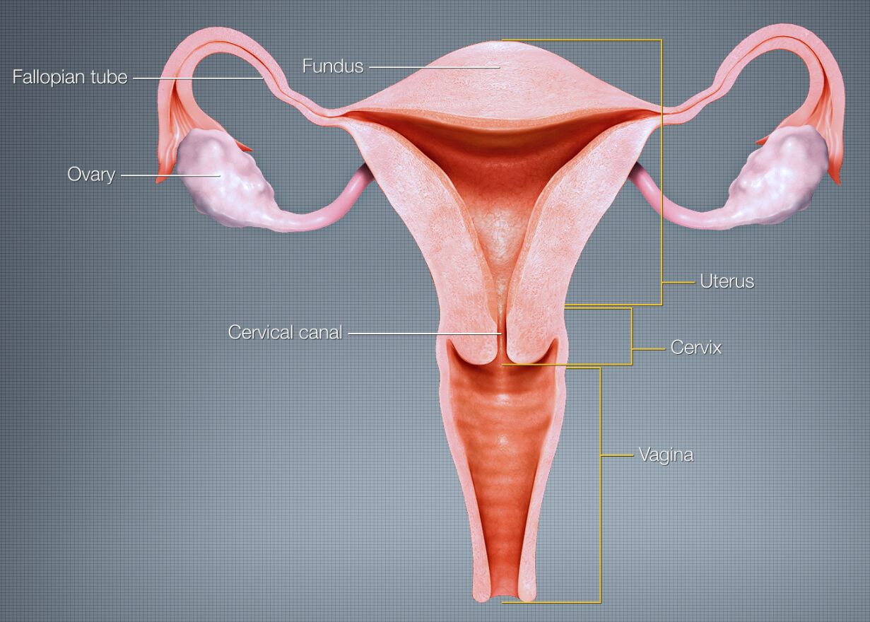
By https://www.scientificanimations.com – https://www.scientificanimations.com/wiki-images/, CC BY-SA 4.0, Link
Fallopian tube The fallopian tubes are paired cylindrical structures extending laterally from the uterine cornua. These specialized tubes measure 10-12 cm in length and contain ciliated epithelium that facilitates egg transport while providing an optimal environment for fertilization.
Ovary The ovaries are paired endocrine organs located in the lateral pelvic cavity. These essential structures produce eggs and hormones including estrogen (E2), progesterone (P4), inhibin A/B, and small amounts of androgens, while containing approximately 1-2 million primordial follicles at birth.
Fundus The fundus is the superior portion of the uterus above the entrance points of the fallopian tubes. This muscular region demonstrates remarkable elasticity and expansion capacity during pregnancy, while maintaining rich vascularization through arcuate and radial arteries.
Uterus The uterus is a thick-walled, pear-shaped organ situated in the pelvic cavity. This remarkable organ can expand from 60-70g to 1000g during pregnancy while maintaining coordinated contractility through its specialized smooth muscle architecture.
Cervical canal The cervical canal is the central passageway through the cervix connecting the uterine cavity to the vagina. This canal contains specialized mucus-secreting glands and undergoes significant changes in response to hormonal fluctuations throughout the menstrual cycle.
Cervix The cervix is the lower cylindrical portion of the uterus connecting to the vagina. This dynamic structure measures 2.5-3 cm in length and contains predominantly collagen tissue that enables significant remodeling during pregnancy and childbirth.
Vagina The vagina is a fibromuscular canal extending from the vulva to the cervix. This highly elastic organ measures 7-9 cm in length and maintains a complex microbiological environment with a pH of 3.8-4.5 through the presence of Lactobacillus species.
The Female Reproductive System: A Comprehensive Guide
Anatomical Organization
The female reproductive system demonstrates precise architectural organization essential for its multiple functions. Each component maintains specific spatial relationships that ensure optimal reproductive capability. The sagittal view reveals crucial anatomical relationships between reproductive organs.
Upper Reproductive Tract
Tubo-ovarian Complex
The fallopian tubes and ovaries form a functional unit essential for reproduction. This complex demonstrates sophisticated neurovascular connections and ligamentous support that enable coordinated function during the reproductive cycle.
The complex consists of:
- Mesosalpinx
- Mesovarium
- Suspensory ligaments
- Neurovascular bundles
Uterine Architecture
The uterus demonstrates three distinct tissue layers:
- Endometrium: 2-8 mm thickness
- Myometrium: 15-20 mm thickness
- Perimetrium: <1 mm thickness
Lower Reproductive Tract
Cervical Structure
The cervix contains:
- Endocervical canal
- Transformation zone
- External os
- Internal os
This region undergoes cyclic changes in response to:
- Estrogen levels
- Progesterone levels
- Local immune factors
- pH changes
Vaginal Anatomy
The vaginal wall consists of:
- Mucosa
- Muscularis
- Adventitia
Clinical Applications
Diagnostic Considerations
Modern imaging techniques include:
- Transvaginal ultrasound (resolution 0.5-1 mm)
- MRI (resolution 1-2 mm)
- CT for specific conditions
- Hysterosalpingography
Surgical Implications
Understanding anatomical relationships is crucial for:
- Hysterectomy approaches
- Fertility procedures
- Oncologic surgery
- Reconstructive procedures
Reproductive Endocrinology
Key hormonal interactions include:
- GnRH pulses (every 90-120 minutes)
- FSH/LH regulation
- Estradiol feedback mechanisms
- Progesterone modulation
Future Perspectives
Emerging technologies focus on:
- 3D organ printing
- Tissue engineering
- Minimally invasive surgery
- Reproductive biotechnology
- Female Reproductive System: A Sagittal View Anatomical Guide
- Understanding Female Reproductive Organ Relationships: Clinical Guide
- Comprehensive Analysis of Female Reproductive Anatomy
- Female Reproductive System: From Structure to Function
- Anatomical Guide to Female Reproductive Organs: Clinical Perspective

