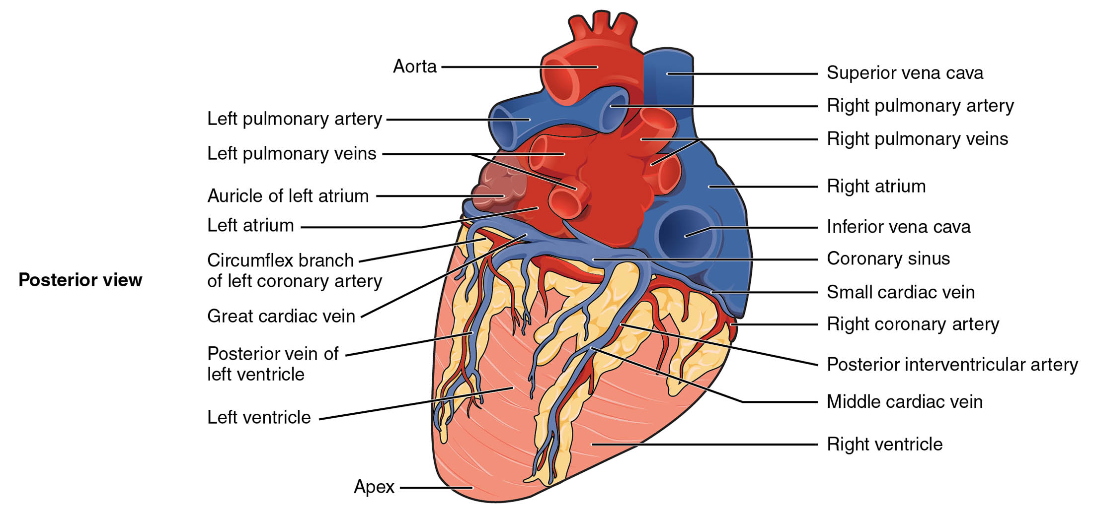
The posterior view of the heart provides a unique perspective on its external structure, revealing key vessels and chambers critical to circulation. This diagram showcases the heart’s back side, highlighting the arteries, veins, and anatomical landmarks that support its function within the thoracic cavity. Studying this image offers valuable insights into the heart’s complex network and its role in sustaining life.
Labelled Parts Explanation
- Aorta The aorta is the largest artery, carrying oxygenated blood from the left ventricle to the systemic circulation. Its posterior position in this view reflects its role in distributing blood to the body.
- Left pulmonary artery The left pulmonary artery transports deoxygenated blood from the right ventricle to the left lung for oxygenation. It branches within the lung to facilitate gas exchange.
- Left pulmonary veins The left pulmonary veins return oxygenated blood from the left lung to the left atrium. They are essential for preparing blood for systemic distribution.
- Auricle of left atrium The auricle of the left atrium is a small, ear-like appendage that increases the atrium’s volume. It receives oxygenated blood from the pulmonary veins.
- Left atrium The left atrium collects oxygenated blood from the pulmonary veins and pumps it into the left ventricle. Its posterior location is key for receiving blood from the lungs.
- Circumflex branch of left coronary artery The circumflex branch of the left coronary artery supplies oxygenated blood to the left atrium and posterior wall of the left ventricle. It is vital for left heart perfusion.
- Great cardiac vein The great cardiac vein drains deoxygenated blood from the anterior heart surface into the coronary sinus. It plays a significant role in cardiac venous return.
- Posterior vein of left ventricle The posterior vein of the left ventricle drains deoxygenated blood from the posterior left ventricular wall. It contributes to the coronary sinus drainage system.
- Left ventricle The left ventricle pumps oxygenated blood into the aorta for systemic circulation. Its thick muscular walls generate the high pressure needed for body-wide distribution.
- Superior vena cava The superior vena cava returns deoxygenated blood from the upper body to the right atrium. Its posterior position aids in venous return to the heart.
- Right pulmonary artery The right pulmonary artery carries deoxygenated blood from the right ventricle to the right lung. It supports pulmonary circulation for oxygenation.
- Right pulmonary veins The right pulmonary veins transport oxygenated blood from the right lung to the left atrium. They ensure efficient blood flow into the systemic circuit.
- Right atrium The right atrium receives deoxygenated blood from the superior and inferior vena cava. It contracts to push blood into the right ventricle.
- Inferior vena cava The inferior vena cava brings deoxygenated blood from the lower body to the right atrium. It complements the superior vena cava in venous return.
- Coronary sinus The coronary sinus collects deoxygenated blood from the heart’s veins, including the great and middle cardiac veins. It empties into the right atrium, supporting cardiac drainage.
- Small cardiac vein The small cardiac vein drains deoxygenated blood from the posterior heart surface. It contributes to the coronary sinus, aiding venous return.
- Right coronary artery The right coronary artery supplies oxygenated blood to the right atrium, right ventricle, and parts of the left ventricle. It is crucial for the heart’s own blood supply.
- Posterior interventricular artery The posterior interventricular artery, a branch of the right coronary artery, supplies blood to the posterior heart and interventricular septum. It ensures adequate posterior perfusion.
- Middle cardiac vein The middle cardiac vein drains deoxygenated blood from the posterior interventricular septum. It empties into the coronary sinus, supporting cardiac venous flow.
- Right ventricle The right ventricle pumps deoxygenated blood into the pulmonary arteries for oxygenation. Its thinner walls are suited to the lower pressure of pulmonary circulation.
- Right pulmonary veins The right pulmonary veins return oxygenated blood from the right lung to the left atrium. They are vital for systemic blood flow preparation.
- Apex The apex is the pointed lower end of the heart, resting on the diaphragm. It marks the inferior tip where the left ventricle extends downward.
- Superior vena cava The superior vena cava returns deoxygenated blood from the upper body to the right atrium. Its posterior position aids in venous return to the heart.
- Right marginal artery The right marginal artery is a branch of the right coronary artery, supplying blood to the lower margin of the right ventricle. It ensures adequate perfusion of this region.
Anatomical Overview of the Heart’s Posterior Features
The posterior view reveals the heart’s back side, showcasing its vascular and chamber anatomy. This perspective highlights the interplay between arteries, veins, and cardiac structures.
- The aorta and pulmonary arteries emerge from the heart’s base, directing blood to the body and lungs.
- The left atrium and right atrium receive blood, with the auricle of left atrium enhancing capacity.
- The left ventricle and right ventricle pump blood, with veins like the great cardiac vein and middle cardiac vein ensuring drainage.
- The coronary sinus serves as a central collector for deoxygenated blood from the heart.
This layout supports the heart’s dual role in pulmonary and systemic circulation.
Major Arteries in the Posterior View
Arteries in the posterior view are critical for supplying the heart and lungs. This diagram outlines their paths and functions.
- The circumflex branch of left coronary artery and posterior interventricular artery nourish the left and posterior heart regions.
- The right coronary artery extends to supply the right heart and septum.
- The left pulmonary artery and right pulmonary artery deliver deoxygenated blood to the lungs.
- The aorta serves as the main conduit for systemic circulation from the left ventricle.
These arteries ensure the heart and lungs receive adequate oxygenation.
Key Veins in Posterior Circulation
Veins are essential for returning blood to the heart in this view. Their placement supports efficient drainage.
- The superior vena cava and inferior vena cava bring deoxygenated blood to the right atrium.
- The left pulmonary veins and right pulmonary veins deliver oxygenated blood to the left atrium.
- The great cardiac vein, middle cardiac vein, and small cardiac vein drain the heart into the coronary sinus.
- The posterior vein of left ventricle supports left ventricular drainage.
This venous network maintains cardiac and pulmonary blood flow.
Chambers and Their Posterior Anatomy
The heart’s chambers are visible from the back, each with specific roles. This anatomy supports their pumping functions.
- The left atrium and right atrium act as receiving chambers, with the auricle of left atrium increasing volume.
- The left ventricle and right ventricle pump blood, with the apex marking the heart’s lower end.
- The left ventricle’s thick walls drive systemic circulation.
- The right ventricle’s thinner structure suits pulmonary pressure.
These features reflect the heart’s adaptation to its circulatory demands.
Clinical Relevance of Posterior Heart Structures
The posterior anatomy aids in diagnosing and treating heart conditions. These structures are key clinical landmarks.
- Blockage of the posterior interventricular artery can lead to posterior wall infarction.
- Dilation of the pulmonary arteries may indicate pulmonary hypertension.
- The coronary sinus is a target for procedures like cardiac resynchronization therapy.
- Veins like the great cardiac vein are critical in coronary angiography.
This knowledge supports effective cardiovascular care and intervention.
Conclusion
The external anatomy of the heart in this posterior view offers a detailed perspective on its vascular and structural components. By understanding the roles of the coronary arteries, pulmonary vessels, and cardiac chambers, one gains insight into the heart’s circulatory efficiency. This knowledge provides a solid foundation for exploring cardiovascular physiology and addressing related health challenges, encouraging further study of the heart’s intricate design and clinical importance.

