The heart’s external anatomy offers a fascinating glimpse into its structure and function, visible once the pericardium is removed. This anterior view diagram highlights the major arteries, veins, and chambers that facilitate blood circulation, providing a clear understanding of the heart’s layout. Examining this image reveals the intricate network that sustains the body’s cardiovascular system.
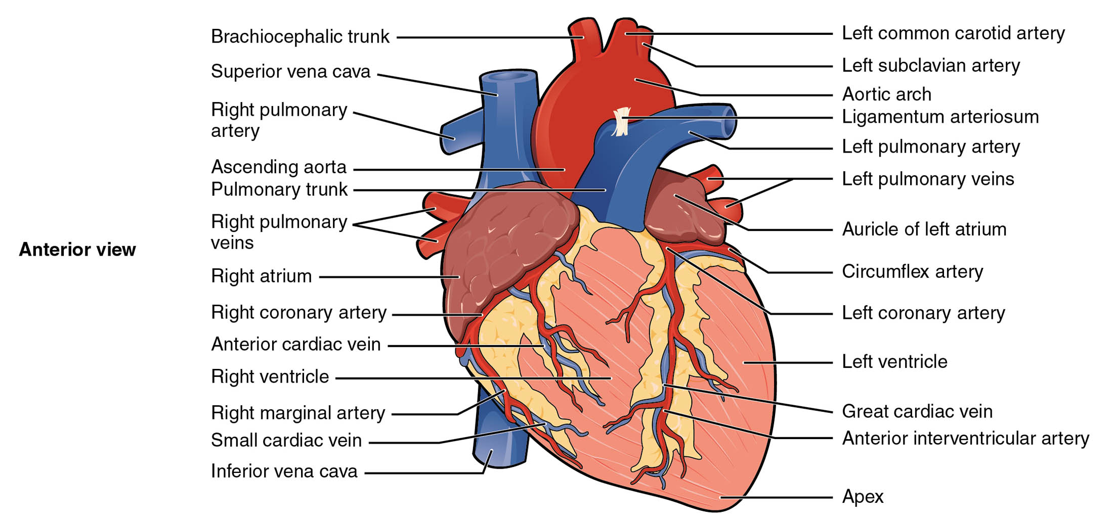
Labelled Parts Explanation
- Brachiocephalic trunk The brachiocephalic trunk is a large artery that branches from the aortic arch, supplying blood to the right arm and head. It divides into the right subclavian and right common carotid arteries.
- Superior vena cava The superior vena cava is a major vein that returns deoxygenated blood from the upper body to the right atrium. It plays a crucial role in the venous return to the heart.
- Right pulmonary artery The right pulmonary artery carries deoxygenated blood from the right ventricle to the right lung for oxygenation. It branches within the lung to facilitate gas exchange.
- Ascending aorta The ascending aorta is the initial segment of the aorta, rising from the left ventricle to distribute oxygenated blood. It gives rise to the coronary arteries that nourish the heart muscle.
- Pulmonary trunk The pulmonary trunk transports deoxygenated blood from the right ventricle to the lungs, dividing into the left and right pulmonary arteries. It is a key component of pulmonary circulation.
- Right pulmonary veins The right pulmonary veins return oxygenated blood from the right lung to the left atrium. They ensure the blood is ready for systemic circulation.
- Right atrium The right atrium receives deoxygenated blood from the superior and inferior vena cava. It contracts to push blood into the right ventricle.
- Right coronary artery The right coronary artery supplies oxygenated blood to the right atrium, right ventricle, and parts of the left ventricle. It is vital for the heart’s own blood supply.
- Anterior cardiac vein The anterior cardiac vein drains deoxygenated blood from the anterior surface of the heart. It empties into the right atrium, supporting cardiac venous return.
- Right ventricle The right ventricle pumps deoxygenated blood into the pulmonary trunk for oxygenation in the lungs. Its thinner walls are adapted to the lower pressure of pulmonary circulation.
- Right marginal artery The right marginal artery is a branch of the right coronary artery, supplying blood to the lower margin of the right ventricle. It ensures adequate perfusion of this region.
- Small cardiac vein The small cardiac vein drains deoxygenated blood from the posterior heart surface. It contributes to the coronary sinus, aiding venous return.
- Inferior vena cava The inferior vena cava returns deoxygenated blood from the lower body to the right atrium. It complements the superior vena cava in the venous system.
- Left common carotid artery The left common carotid artery arises from the aortic arch, delivering oxygenated blood to the left side of the head and neck. It branches into internal and external carotid arteries.
- Left subclavian artery The left subclavian artery supplies oxygenated blood to the left arm and parts of the chest wall. It originates from the aortic arch, supporting upper limb circulation.
- Aortic arch The aortic arch is the curved portion of the aorta that connects the ascending and descending aorta. It gives rise to major arteries supplying the head, neck, and arms.
- Ligamentum arteriosum The ligamentum arteriosum is a remnant of the ductus arteriosus, connecting the pulmonary artery to the aorta in fetal life. It becomes a fibrous band after birth.
- Left pulmonary artery The left pulmonary artery carries deoxygenated blood from the right ventricle to the left lung. It supports oxygenation through pulmonary circulation.
- Left pulmonary veins The left pulmonary veins return oxygenated blood from the left lung to the left atrium. They are essential for systemic blood flow.
- Auricle of left atrium The auricle of the left atrium is a small, ear-like extension that increases the atrium’s capacity. It receives oxygenated blood from the pulmonary veins.
- Circumflex artery The circumflex artery is a branch of the left coronary artery, supplying blood to the left atrium and posterior wall of the left ventricle. It is critical for left heart perfusion.
- Left coronary artery The left coronary artery arises from the ascending aorta, supplying oxygenated blood to the left atrium, left ventricle, and interventricular septum. It is vital for left heart function.
- Left ventricle The left ventricle pumps oxygenated blood into the ascending aorta for systemic circulation. Its thick muscular walls generate high pressure to support body-wide distribution.
- Great cardiac vein The great cardiac vein drains deoxygenated blood from the anterior heart surface into the coronary sinus. It plays a key role in cardiac venous drainage.
- Anterior interventricular artery The anterior interventricular artery, also called the left anterior descending artery, supplies blood to the anterior wall of the heart and interventricular septum. It is crucial for heart muscle oxygenation.
- Apex The apex is the pointed lower end of the heart, resting on the diaphragm. It marks the inferior tip where the left ventricle extends downward.
Anatomical Overview of the Heart’s External Features
The external anatomy of the heart reveals its complex vascular network. This anterior view highlights the arteries and veins that ensure blood flow to and from the heart.
- The ascending aorta and pulmonary trunk emerge from the heart’s base, directing blood to the body and lungs.
- The right atrium and right ventricle handle deoxygenated blood, while the left ventricle manages oxygenated blood.
- The right coronary artery and left coronary artery provide the heart’s own blood supply, branching into smaller vessels like the anterior interventricular artery.
- Veins such as the great cardiac vein and small cardiac vein return deoxygenated blood to the right atrium via the coronary sinus.
This layout supports the heart’s role as a dual pump, sustaining both pulmonary and systemic circulation.
Major Arteries of the Heart
Arteries are the highways of oxygenated blood delivery to the heart and body. This diagram showcases their critical pathways.
- The left coronary artery and right coronary artery originate from the ascending aorta, feeding the heart muscle.
- The anterior interventricular artery and circumflex artery branch from the left coronary artery, supplying the left heart.
- The right marginal artery ensures blood flow to the right ventricle’s margin.
- The aortic arch gives rise to the brachiocephalic trunk, left common carotid artery, and left subclavian artery.
These arteries are essential for maintaining cardiac function and upper body circulation.
Key Veins in Cardiac Circulation
Veins play a vital role in returning deoxygenated blood to the heart. This view highlights their strategic placement.
- The superior vena cava and inferior vena cava deliver blood to the right atrium from the upper and lower body.
- The right pulmonary veins and left pulmonary veins bring oxygenated blood from the lungs to the left atrium.
- The great cardiac vein and small cardiac vein drain the heart’s anterior and posterior surfaces.
- The anterior cardiac vein provides an additional drainage route to the right atrium.
This venous network ensures efficient blood return for reoxygenation.
Chambers and Their External Features
The heart’s chambers are visible externally, each with distinct roles. This anatomy supports their pumping functions.
- The right atrium and left atrium receive blood, with the auricle of left atrium expanding capacity.
- The right ventricle and left ventricle pump blood, with the apex marking the heart’s lower tip.
- The left ventricle’s thick walls reflect its role in systemic circulation.
- The right ventricle’s thinner structure suits pulmonary pressure needs.
These features highlight the heart’s adaptability to its dual circulatory demands.
Clinical Significance of Heart Anatomy
Understanding the external heart anatomy aids in diagnosing and treating cardiovascular conditions. The labeled structures are key landmarks.
- Blockage of the left coronary artery or anterior interventricular artery can lead to myocardial infarction.
- Dilation of the pulmonary trunk may indicate pulmonary hypertension.
- The ligamentum arteriosum is a remnant assessed in congenital heart disease evaluations.
- Veins like the great cardiac vein are critical in cardiac catheterization procedures.
This knowledge supports effective clinical interventions and heart health management.
Conclusion
The external anatomy of the heart, as depicted in this anterior view, provides a detailed map of its vascular and structural components. By exploring the roles of the coronary arteries, pulmonary vessels, and cardiac chambers, one gains a deeper appreciation for the heart’s role in circulation. This understanding serves as a foundation for studying cardiovascular physiology and addressing related health issues, encouraging further exploration of the heart’s remarkable design and function.

