The human brain’s ability to process visual information relies on intricate pathways that transform raw sensory input into meaningful perceptions. This diagram illustrates the division of visual processing into the ventral and dorsal streams, originating from the occipital lobe and extending into the temporal and parietal lobes, respectively, offering a glimpse into how we understand “what” and “where” in our environment.
Labels in the Diagram
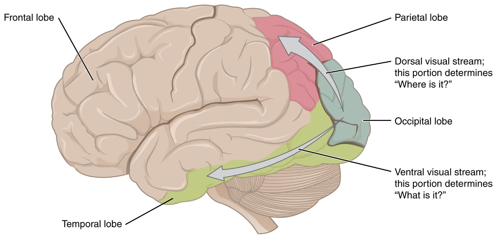
Frontal Lobe The frontal lobe is located at the front of the brain and plays a key role in higher cognitive functions such as decision-making and planning. In the context of visual processing, it integrates information from the dorsal stream to guide spatial awareness and action.
Parietal Lobe The parietal lobe, situated near the top and back of the brain, is critical for processing sensory input and spatial orientation. It houses part of the dorsal visual stream, which helps determine the location of objects in space.
Dorsal Visual Stream The dorsal visual stream, often referred to as the “where” pathway, extends from the occipital lobe into the parietal lobe. This stream is responsible for spatial awareness, motion detection, and coordinating movements based on visual cues.
Occipital Lobe The occipital lobe, located at the rear of the brain, contains the primary visual cortex where initial visual processing occurs. It serves as the starting point for both the dorsal and ventral visual streams.
Temporal Lobe The temporal lobe, found on the sides of the brain, is involved in memory and auditory processing. It hosts the ventral visual stream, which focuses on object recognition and identification.
Ventral Visual Stream The ventral visual stream, known as the “what” pathway, travels from the occipital lobe into the temporal lobe. This stream is essential for identifying objects, recognizing faces, and interpreting visual details.
Anatomy of the Visual Processing Pathways
The visual system begins with the eyes, where light is converted into electrical signals by photoreceptors in the retina. These signals travel via the optic nerve to the occipital lobe, where the primary visual cortex processes basic features like edges and motion.
- Photoreceptors: Rods and cones in the retina detect light intensity and color, initiating the visual signal.
- Optic Nerve: Transmits signals from the retina to the brain, with fibers partially crossing at the optic chiasm.
- Primary Visual Cortex: Located in the occipital lobe, it analyzes orientation and spatial frequency of visual input.
From here, visual information splits into two distinct pathways, each serving a unique purpose in perception.
The Dorsal Visual Stream: The “Where” Pathway
The dorsal visual stream processes spatial relationships and guides movement. This pathway extends from the occipital lobe to the parietal lobe, working closely with the frontal lobe for action planning.
- Spatial Awareness: Enables locating objects in three-dimensional space, crucial for navigation.
- Motion Detection: Neurons here respond to moving stimuli, aiding in tracking objects.
- Motor Coordination: Links visual input to motor responses, such as reaching for an item.
Damage to this stream, as seen in conditions like optic ataxia, can impair the ability to accurately reach for objects, despite intact object recognition.
The Ventral Visual Stream: The “What” Pathway
The ventral visual stream focuses on object identification and recognition. Originating in the occipital lobe, it projects into the temporal lobe, where complex visual features are analyzed.
- Object Recognition: Identifies shapes, colors, and patterns to distinguish objects like a cup or a face.
- Face Perception: Specialized areas, such as the fusiform face area, process facial features.
- Memory Integration: Connects visual input with stored memories for context, like recognizing a friend.
Lesions in this pathway, such as in prosopagnosia, can lead to difficulty recognizing faces, highlighting its role in visual memory.
Integration of Visual Streams in Daily Life
Both streams work together to create a cohesive visual experience. The dorsal visual stream helps us navigate a room, while the ventral visual stream allows us to recognize furniture and decor.
- Everyday Tasks: Driving requires spatial judgment from the dorsal stream and object identification from the ventral stream.
- Learning Environments: Reading maps relies on spatial skills, while reading text depends on object recognition.
- Therapeutic Applications: Understanding these streams aids in rehabilitating patients with visual impairments.
Neuroimaging studies show that these pathways activate differently based on task demands, offering insights into brain plasticity.
Clinical Relevance and Potential Disorders
While the diagram depicts a healthy brain, disruptions in these streams can lead to specific deficits. Damage to the parietal lobe might affect spatial tasks, while temporal lobe injuries could impair object recognition.
- Visual Agnosia: Loss of ability to interpret visual information, often linked to ventral stream damage.
- Balint’s Syndrome: Affects spatial attention and reaching, associated with parietal lobe lesions.
- Rehabilitation Strategies: Visual therapy can sometimes compensate for stream-specific deficits.
Early diagnosis through behavioral tests and imaging can guide treatment, emphasizing the importance of these pathways.
Evolutionary Perspective and Future Research
The division into dorsal and ventral visual streams likely evolved to enhance survival, with spatial skills aiding hunting and object recognition supporting foraging. Modern research continues to explore how these streams adapt to technology, like virtual reality.
- Evolutionary Advantage: Forward-facing eyes in primates support binocular vision, enhancing both streams.
- Technological Impact: Augmented reality leverages these pathways for immersive experiences.
- Ongoing Studies: Investigating neural plasticity may improve treatments for visual pathway disorders.
Advances in functional MRI provide detailed maps of these streams, promising better therapeutic outcomes.
In conclusion, the ventral and dorsal visual streams represent a remarkable collaboration within the brain, turning light into a rich tapestry of spatial and object-based understanding. This diagram serves as a valuable tool for exploring the intricacies of vision, offering a foundation for both clinical practice and scientific inquiry.

