The respiratory zone is a fascinating part of the human body where oxygen and carbon dioxide are exchanged, a process vital for sustaining life. This article provides an in-depth look at the alveoli and their anatomical features as depicted in the provided diagram, offering insights into their structure and function for a better understanding of respiratory health.
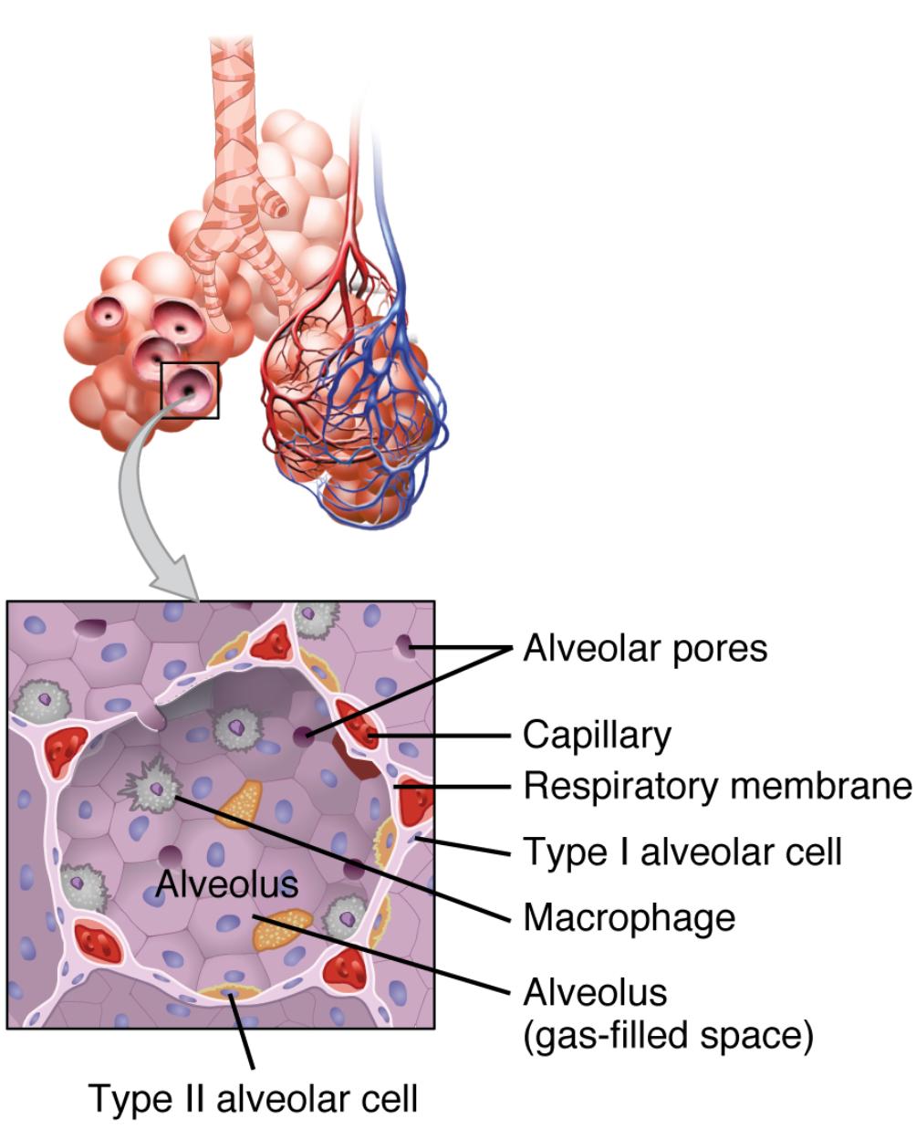
Alveolar pores: These small openings connect adjacent alveoli, allowing air pressure to equalize and providing alternative air pathways. They play a key role in maintaining ventilation, especially when some airways are obstructed.
Capillary: These tiny blood vessels envelop the alveoli, facilitating the exchange of oxygen and carbon dioxide with a wall thickness of just 0.5-1 micrometer. Their extensive network ensures efficient gas diffusion into the bloodstream.
Respiratory membrane: This ultra-thin layer, comprising the alveolar wall and capillary endothelium, enables rapid gas exchange due to its minimal thickness. Its integrity is crucial for the effective transfer of oxygen into the blood and removal of carbon dioxide.
Type I alveolar cell: These flat, squamous cells line most of the alveoli, offering a thin barrier for gas exchange with a thickness of about 0.1-0.5 micrometers. They are essential for the diffusion process without adding significant resistance.
Macrophage: These immune cells roam the alveoli, clearing dust, bacteria, and debris to protect lung health. Their presence helps prevent infections and maintains a clean environment for gas exchange.
Alveolus (gas-filled space): This air-filled sac is the site where oxygen enters the blood and carbon dioxide is expelled, supported by its elastic structure. Its large surface area, roughly 70-100 square meters in adults, enhances respiratory efficiency.
Type II alveolar cell: These cuboidal cells secrete surfactant to reduce surface tension and prevent alveolar collapse during exhalation. They also contribute to repairing the alveolar lining when damage occurs.
Anatomical Breakdown of the Alveoli
The alveoli serve as the cornerstone of the respiratory system, where the exchange of gases takes place with remarkable efficiency. This section breaks down the key components that make this process possible.
- The alveolus (gas-filled space) is a tiny sac filled with air, designed to maximize the surface area for gas exchange.
- Surrounding each alveolus, capillaries form a dense network, ensuring close contact for oxygen to diffuse into the blood.
- Alveolar pores act as safety valves, equalizing pressure and providing backup airways when needed.
- Type I alveolar cells provide a thin, specialized lining that supports the rapid movement of gases across the respiratory membrane.
Cellular Contributions to Respiratory Function
The cells within the alveoli play a critical role in maintaining respiratory health and efficiency. This part highlights their specific functions and importance.
- Macrophages patrol the alveolar space, engulfing pathogens and particles to keep the lungs free from infection.
- Type II alveolar cells produce surfactant, a lipid-protein mixture that prevents the alveoli from collapsing after each breath.
- Type I alveolar cells, with their thin structure, are optimized for gas diffusion, making up about 95% of the alveolar surface.
- The respiratory membrane, supported by these cells, ensures a seamless exchange of oxygen and carbon dioxide.
The Role of Blood Vessels and Membranes
Blood vessels and membranes are integral to the gas exchange process, working in harmony with alveolar structures. This section explores their contributions in detail.
- Capillaries surrounding the alveoli are so thin that gases can diffuse across in a fraction of a second, delivering oxygen to red blood cells.
- The respiratory membrane’s thinness, combined with its large surface area, allows for efficient gas transfer under normal breathing conditions.
- These structures adapt to increased demand during physical activity, enhancing oxygen delivery to tissues.
- The close proximity of capillaries to alveoli ensures that deoxygenated blood is quickly refreshed with each breath.
Conclusion
The intricate design of the respiratory zone, particularly the alveoli, showcases the body’s remarkable ability to sustain life through efficient gas exchange. From the protective role of macrophages to the surfactant production by Type II alveolar cells, each component works together to ensure optimal respiratory function. Exploring these structures provides valuable insights into the anatomy and physiology of the lungs, fostering a deeper appreciation for this vital system.

