Delve into the external anatomy of the human heart through detailed dissections, revealing the key chambers and major blood vessels that orchestrate life-sustaining circulation. This article provides an in-depth look at structures like the aorta, pulmonary trunk, and the right and left ventricles, crucial for understanding cardiac function. Gain valuable insights into the heart’s complex design and its vital role in the cardiovascular system.
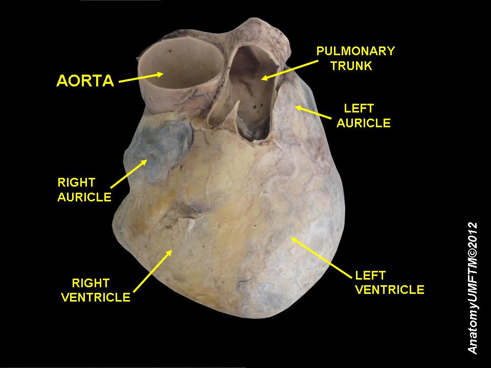
Aorta: The aorta is the largest artery in the human body, originating from the left ventricle of the heart and extending down to the abdomen. It is responsible for distributing oxygenated blood from the heart to all parts of the body, functioning as the primary conduit of systemic circulation.
Pulmonary Trunk: The pulmonary trunk is a large artery that arises from the right ventricle of the heart, bifurcating into the right and left pulmonary arteries. It carries deoxygenated blood to the lungs, where it will undergo gas exchange to become oxygenated.
Left Auricle: The left auricle is a small, muscular pouch that projects from the anterior surface of the left atrium. It functions as an expansion chamber for the left atrium, allowing it to hold additional blood before it is pumped into the left ventricle.
Right Auricle: Similar to its left counterpart, the right auricle is a small, conical muscular pouch extending from the right atrium. It serves as an accessory blood reservoir for the right atrium, temporarily holding deoxygenated blood returning from the body before it enters the main atrial chamber.
Right Ventricle: The right ventricle is one of the two lower chambers of the heart, responsible for pumping deoxygenated blood into the pulmonary trunk, which then carries it to the lungs. It has thinner muscular walls compared to the left ventricle, as it pumps blood over a shorter distance and against lower pressure.
Left Ventricle: The left ventricle is the most muscular chamber of the heart, responsible for pumping oxygenated blood into the aorta, from where it is distributed throughout the entire body. Its thick walls generate the high pressure needed to ensure systemic circulation and meet the metabolic demands of all tissues.
The human heart is an indispensable organ, a tireless pump that circulates blood throughout the body, delivering oxygen and nutrients while removing metabolic waste products. Its intricate anatomical design ensures the efficient separation of oxygenated and deoxygenated blood, a hallmark of the mammalian double circulatory system. Detailed anatomical dissections, like the one shown, offer invaluable insights into the external features of this vital organ, highlighting its four chambers and the major blood vessels that either arise from or enter it. Understanding these structures is foundational for comprehending both normal cardiac function and the pathophysiology of cardiovascular diseases.
The heart is strategically located in the mediastinum, slightly to the left of the midline in the thoracic cavity, nestled between the lungs. Its external surface is characterized by various grooves, or sulci, that delineate the boundaries of its four chambers: two atria and two ventricles. The atria are the superior receiving chambers, while the ventricles are the inferior pumping chambers. Emerging from the base of the heart are the great vessels—the aorta and the pulmonary trunk—which are critical for distributing blood to the systemic and pulmonary circuits, respectively. The auricles, small ear-like appendages, are visible extensions of the atria, acting as reserve chambers.
The robust muscular walls of the heart, particularly the ventricles, are crucial for generating the pressure required to propel blood. The left ventricle, needing to pump blood to the entire body, possesses a significantly thicker myocardial wall than the right ventricle, which only pumps blood to the lungs. This structural difference reflects their distinct functional demands. The precise positioning and interconnections of these chambers and vessels ensure a unidirectional flow of blood, preventing mixing of oxygenated and deoxygenated blood, which is essential for maintaining high metabolic rates and supporting complex physiological processes.
Key external anatomical features of the heart include:
- Four chambers: Right atrium, left atrium, right ventricle, left ventricle.
- Great vessels: Aorta and pulmonary trunk.
- Auricles: Appendages of the atria.
- Coronary sulcus: Separates atria from ventricles.
- Interventricular sulci: Separate the right and left ventricles.
In conclusion, the meticulous anatomical dissection of the human heart provides a profound appreciation for its complex external architecture. The precise arrangement of the aorta, pulmonary trunk, auricles, and the distinct right and left ventricle chambers underscores the heart’s specialized role as the central engine of the cardiovascular system. A comprehensive understanding of these structures is paramount for medical professionals, enabling them to diagnose and treat a myriad of cardiac conditions, ultimately contributing to the preservation of life and well-being.

