Exploring the External Nose: Anatomy and Structure

Date:

The external nose is a prominent facial feature that plays a vital role in respiration and olfaction, as detailed in the provided diagram. This article delves into the anatomical components illustrated, offering a thorough understanding of its structure and function. By examining these elements, one can appreciate the nose’s significance in both aesthetic and physiological contexts.

Exploring the External Nose: Anatomy and Structure

Nasal bone: These paired bones form the upper bridge of the nose, providing structural support and protection. They articulate with the frontal bone and maxilla, contributing to the nose’s rigidity.

Nasal cartilage: This flexible tissue, including the septal and alar cartilages, shapes the lower and middle portions of the nose. It allows for slight movement and resilience while maintaining the nasal airway.

- Advertisement -

Recommended Study Resource

Gray's Anatomy: The Anatomical Basis of Clinical Practice

Enhance your anatomical knowledge with Gray's Anatomy: The Anatomical Basis of Clinical Practice. This authoritative text offers in-depth insights and illustrations, perfect for medical students and practitioners aiming for clinical excellence.

Shop Now on Amazon

At AnatomyNote.com, we offer free resources on anatomy, pathology, and pediatric medicine for medical students and professionals. Purchasing through our Amazon links, like Gray's Anatomy, supports our server costs and content creation at no additional cost to you.

Disclosure: As an Amazon Associate, we earn a commission from qualifying purchases.

Disclosure: As an Amazon Associate, we earn a commission from qualifying purchases at no extra cost to you.

Nasal septum: This vertical partition, composed of cartilage and bone, divides the nasal cavity into two nostrils. It ensures proper airflow and supports the overall nasal framework.

Ala nasi: These wing-like structures form the outer edges of the nostrils, made of cartilage and soft tissue. They help regulate airflow and can dilate or constrict during breathing.

Nostril (Naris): This external opening of the nasal cavity allows air to enter and exit the respiratory system. Each nostril is lined with hair and mucus to filter incoming air.

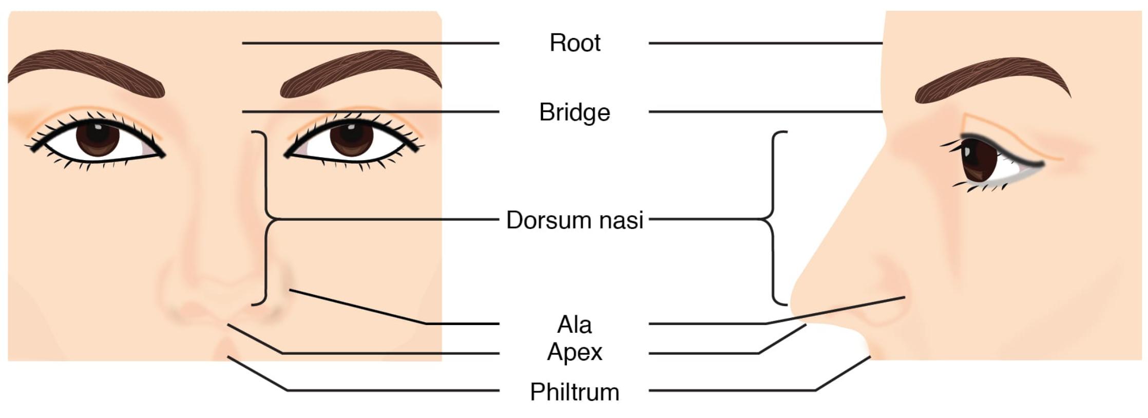
Philtrum: This midline groove runs from the nasal septum to the upper lip, marking the fusion line of facial tissues during embryonic development. It adds to the nose’s aesthetic contour.

- Advertisement -

Anatomy Flash Cards

Master anatomy with detailed, exam-ready flash cards.

Buy on Amazon

AnatomyNote.com offers free anatomy and pathology resources. Your purchase of Anatomy Flash Cards supports our site at no extra cost.

As an Amazon Associate, we earn from qualifying purchases.

Anatomical Structure of the External Nose

The external nose’s anatomy is a blend of bone and cartilage, designed for both function and form. Each part contributes to its overall effectiveness.

  • The nasal bone provides a sturdy foundation for the upper nose.
  • The nasal cartilage offers flexibility, adapting to facial expressions.
  • The nasal septum ensures symmetrical airflow through both nostrils.
  • The ala nasi supports the nostrils’ shape and airflow regulation.
  • The nostril (naris) serves as the entry point for respiratory and olfactory functions.

Physiological Functions of the External Nose

The external nose performs essential roles in respiration and sensory perception. Its structure supports these critical processes.

  • The nasal bone and nasal cartilage protect the nasal passages from injury.
  • The nasal septum directs airflow, preventing obstruction.
  • The ala nasi adjusts nostril size to optimize breathing efficiency.
  • The nostril (naris) filters dust and pathogens with nasal hair.
  • The philtrum has no direct physiological role but aids in facial recognition.

Clinical Relevance and Health Considerations

While the diagram focuses on anatomy, the external nose’s health is vital for overall well-being. Awareness of potential issues can guide care.

  • Fractures of the nasal bone are common and may require surgical realignment.
  • Deviated nasal septum can cause breathing difficulties, treatable with septoplasty.
  • Infections or injuries to the ala nasi may affect airflow and require antibiotics.
  • Narrow nostril (naris) openings can lead to snoring or sleep apnea.
  • The philtrum’s appearance can be altered by cleft lip, correctable with surgery.

The external nose, as depicted, showcases the harmonious integration of the nasal bone, nasal cartilage, nasal septum, ala nasi, nostril (naris), and philtrum in supporting respiration and olfaction. This anatomical guide provides a foundation for exploring its physiological roles and clinical significance. By studying these structures, one can better understand their importance in maintaining health and addressing related conditions effectively.

We'd be thrilled to have your support!

Your generous contribution through a coffee keeps our passion alive.

Image source:

LEAVE A REPLY

Please enter your comment!
Please enter your name here

Share post:

Popular

spot_imgspot_img

Subscribe

More like this
Related

The Human Knee Anatomy: Understanding Joint Anatomy and Function

The knee joint represents one of the most complex and crucial articulations in the human body, essential for mobility and weight-bearing activities. This detailed anterior view illustration demonstrates the primary anatomical structures that work together to provide stability, flexibility, and smooth movement in the knee joint. Each component plays a vital role in maintaining proper knee function and preventing injury.

Diagram of Spongy Bone: Exploring Anatomical Structure and Function

The spongy bone, also known as cancellous bone, is a vital component of the skeletal system, characterized by its porous structure that supports both mechanical and physiological roles. This medical image provides a detailed diagram of spongy bone, showcasing its trabecular framework and the presence of red marrow within its spaces. By examining these labeled components, we can understand how spongy bone contributes to lightweight strength, shock absorption, and blood cell production, making it essential for skeletal health and overall body function.

Laryngeal cartilages anatomical structure

The laryngeal cartilages form an intricate structural framework that is essential for voice production, airway protection, and breathing. This detailed anatomical illustration showcases the various cartilages and structures that comprise the larynx, including the hyoid bone, thyroid cartilage, cricoid cartilage, and epiglottis. Each component plays a vital role in maintaining proper laryngeal function and protecting the airway during swallowing.

Exploring the Nasal Septum: Anatomical Structure and Function

The nasal septum, a crucial midline structure, divides the nasal cavity into two symmetrical halves, facilitating efficient airflow and olfaction. Formed by the perpendicular plate of the ethmoid bone, the vomer bone, and the septal cartilage, it integrates with surrounding bones like the frontal bone and maxilla, as depicted in a sagittal section. This article delves into the anatomy of the nasal septum, its associated structures, and their physical characteristics, providing a comprehensive resource for understanding nasal anatomy.