Embark on a fascinating journey through the embryological development of the human heart, tracing its transformation from a simple tube to a complex four-chambered organ. This detailed guide utilizes a comprehensive diagram to illustrate the critical stages of heart formation during the first eight weeks of embryonic life, offering insights into the anatomical and physiological foundations of cardiovascular health. Whether you’re delving into the initial blood flow patterns or the partitioning into atria and ventricles, this article provides a clear and engaging overview of this vital process.
Label Introduction
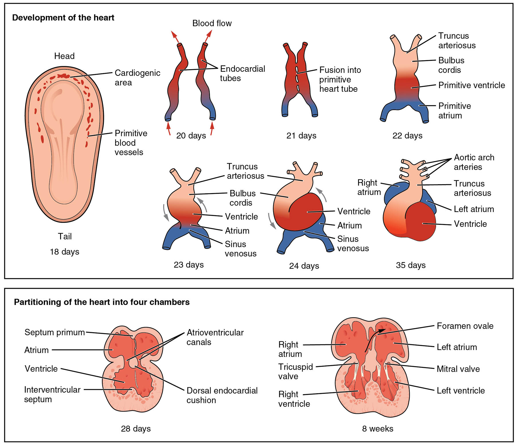
- Cardiogenic area The cardiogenic area is the region in the embryo where the heart begins to form, located in the cranial part of the primitive streak. It becomes the site where primitive blood vessels and the heart tube will develop.
- Primitive blood vessels Primitive blood vessels are the initial network of blood-carrying structures that emerge around the cardiogenic area, facilitating early circulation. These vessels evolve into the complex vascular system supporting the developing heart.
- Endocardial tubes Endocardial tubes are the paired structures that fuse to form the primitive heart tube, marking a key step at around 20 days of development. This fusion sets the stage for the heart’s tubular structure and initial blood flow.
- Fusion into primitive heart tube Fusion into primitive heart tube occurs at approximately 21 days, where the endocardial tubes merge to create a single tubular heart. This tube begins to exhibit rhythmic contractions to circulate blood.
- Truncus arteriosus The truncus arteriosus is the outflow tract of the primitive heart tube, present by 23 days, which will later divide into the aorta and pulmonary artery. It plays a crucial role in directing blood flow from the ventricles.
- Bulbus cordis Bulbus cordis is a segment of the heart tube adjacent to the truncus arteriosus, visible at 23 days, which contributes to the formation of the right ventricle and parts of the outflow tracts. Its development is essential for proper ventricular separation.
- Ventricle The ventricle begins as a dilated portion of the heart tube by 23 days, serving as the pumping chamber that will eventually split into left and right ventricles. It undergoes significant remodeling during heart partitioning.
- Atrium The atrium forms as a distinct region of the heart tube by 23 days, initially receiving blood from the sinus venosus, and will later divide into the left and right atria. This area is critical for coordinating blood entry into the ventricles.
- Sinus venosus Sinus venosus is the inflow tract of the primitive heart at 23 days, collecting blood from the embryonic veins and delivering it to the atrium. It eventually contributes to the formation of the sinoatrial node and parts of the atria.
- Right atrium The right atrium emerges as a defined chamber by 35 days, receiving deoxygenated blood from the body via the superior and inferior vena cava. It plays a key role in the eventual four-chambered heart structure.
- Left atrium The left atrium begins to form by 35 days, receiving oxygenated blood from the lungs via the pulmonary veins, preparing for its role in systemic circulation. Its development is vital for the separation of oxygenated and deoxygenated blood.
- Aortic arches Aortic arches are a series of arterial structures visible by 22 days, which contribute to the formation of the great vessels like the aorta and pulmonary arteries. They undergo complex remodeling to support adult circulation.
- Bulbus cordis This region, noted again at 22 days, further develops into parts of the ventricles and outflow tracts, emphasizing its dual role in heart morphogenesis. Its precise division is crucial for normal heart function.
- Primitive ventricle The primitive ventricle, seen at 22 days, is the early pumping chamber that will differentiate into the left and right ventricles. Its growth and partitioning are fundamental to the heart’s pumping efficiency.
- Primitive atrium The primitive atrium, present at 22 days, is the initial receiving chamber that will split into the left and right atria. It sets the foundation for the heart’s ability to handle dual circulatory systems.
- Left atrium ventricle The left atrium ventricle, observed at 35 days, represents the developing left side of the heart, integrating atrial and ventricular functions. This area will become the left ventricle, pumping oxygenated blood into the aorta.
- Matril valve The mitral valve, forming by 8 weeks, separates the left atrium from the left ventricle, ensuring unidirectional blood flow. Its proper development is essential to prevent backflow and maintain efficient circulation.
- Right ventricle The right ventricle, defined by 8 weeks, pumps deoxygenated blood into the pulmonary artery, completing the right side of the heart. Its formation is critical for pulmonary circulation.
- Tricuspid valve The tricuspid valve, present by 8 weeks, lies between the right atrium and ventricle, preventing backflow during contraction. Its development ensures effective right heart function.
- Foramen ovale The foramen ovale, visible at 8 weeks, is an opening in the atrial septum allowing fetal blood to bypass the lungs. It typically closes after birth, transitioning to adult circulation.
- Septum primum The septum primum, forming at 28 days, is a partition that begins to divide the atrium into left and right sides. It plays a key role in the eventual closure of the foramen ovale.
- Atrioventricular canals Atrioventricular canals, seen at 28 days, are the passages connecting the atria and ventricles, guiding early blood flow. They contribute to the formation of the mitral and tricuspid valves.
- Dorsal endocardial cushion The dorsal endocardial cushion, present at 28 days, is a tissue mass that helps fuse the septum primum and atrioventricular canals. It is vital for the structural integrity of the heart chambers.
- Interventricular septum The interventricular septum, forming at 28 days, begins to separate the ventricles into left and right chambers. Its complete closure is essential for the heart’s four-chambered configuration.
Introduction to Heart Development
The journey of heart development begins early in embryonic life, laying the groundwork for a robust cardiovascular system. This process transforms a simple cardiogenic area into a fully functional four-chambered heart by eight weeks.
- Outlines the initial formation of the cardiogenic area and primitive blood vessels as the starting point of heart development.
- Describes the fusion of endocardial tubes into a primitive heart tube, initiating blood flow.
- Highlights the progressive complexity as the heart adapts to the growing embryo’s needs.
Early Stages of Heart Formation
The heart’s initial development sets the stage for its lifelong role in circulation. From 18 to 22 days, the embryo witnesses rapid changes in heart structure.
- Details the emergence of primitive blood vessels around the cardiogenic area, supporting early circulation.
- Explains how endocardial tubes fuse at 20 days to form the primitive heart tube, enabling rhythmic contractions.
- Notes the appearance of the truncus arteriosus and bulbus cordis by 22 days, marking the outflow and ventricular regions.
Progression to a Tubular Heart
By 23 to 24 days, the heart tube undergoes significant looping and differentiation. This stage is crucial for establishing the basic heart layout.
- Describes the looping of the ventricle and atrium, driven by the sinus venosus inflow.
- Explains the role of the truncus arteriosus in directing blood flow as the heart tube elongates.
- Highlights the bulbus cordis contribution to the ventricular outflow tracts during this phase.
Development of Atria and Ventricles
From 28 days onward, the heart begins partitioning into distinct chambers. This process ensures the separation of oxygenated and deoxygenated blood.
- Discusses the formation of the septum primum and interventricular septum, initiating chamber division.
- Explains how atrioventricular canals guide blood flow and contribute to valve formation.
- Notes the role of the dorsal endocardial cushion in stabilizing the developing septa.
Completion of Four-Chambered Heart
By 8 weeks, the heart achieves its four-chambered structure, ready for postnatal function. This final stage refines the heart’s anatomical and physiological roles.
- Details the separation of the right atrium and left atrium, with the foramen ovale facilitating fetal circulation.
- Describes the development of the right ventricle and left atrium ventricle, supported by the tricuspid valve and mitral valve.
- Highlights the aortic arches remodeling into major arteries, completing the circulatory network.
Physiological Significance of Heart Development
The embryological process ensures the heart can meet the fetus’s and later the newborn’s circulatory demands. Understanding these stages provides insight into congenital anomalies.
- Explains how the sinus venosus evolves into the sinoatrial node, regulating heart rhythm.
- Discusses the primitive atrium and primitive ventricle differentiation into functional chambers.
- Notes the critical role of the bulbus cordis in forming the right ventricle and outflow tracts.
Clinical Relevance of Embryonic Heart Development
Knowledge of heart development aids in identifying and addressing congenital heart defects. Early stages like those involving the truncus arteriosus and aortic arches are prone to malformations.
- Identifies potential issues with septum primum closure leading to atrial septal defects.
- Explains how incomplete interventricular septum formation can cause ventricular septal defects.
- Highlights the importance of proper atrioventricular canals development for valve function.
In conclusion, the embryological development of the human heart is a remarkable process that transforms a simple tube into a sophisticated organ. Each stage, from the cardiogenic area to the fully formed four chambers, builds on the previous one, ensuring the heart can sustain life. This diagram serves as an essential tool for understanding the anatomical and physiological foundations of the cardiovascular system, offering valuable insights into both normal development and potential congenital variations.

