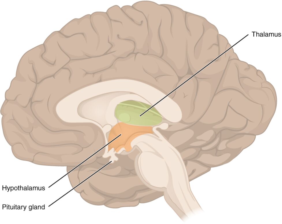
The diencephalon represents a crucial subdivision of the forebrain, encompassing structures that integrate sensory, motor, and autonomic functions. This image depicts the diencephalon in a midsagittal view, highlighting the thalamus, hypothalamus, and pituitary gland, which together form the walls of the third ventricle and play vital roles in relaying information and regulating homeostasis. Delving into their anatomy provides foundational knowledge for understanding neural processing and endocrine control.
Thalamus
The thalamus is a paired, ovoid structure located on either side of the third ventricle, serving as the primary relay station for sensory and motor signals to the cerebral cortex. It processes and filters incoming information from various pathways, ensuring only relevant data reaches higher brain centers for conscious perception and response.
Hypothalamus
The hypothalamus, situated inferior and anterior to the thalamus, acts as a central regulator of autonomic functions, including temperature control, hunger, thirst, and circadian rhythms. It also serves as a link between the nervous and endocrine systems by controlling the pituitary gland through releasing and inhibiting hormones.
Pituitary gland
The pituitary gland, attached to the hypothalamus via the infundibulum, is divided into anterior and posterior lobes that secrete hormones influencing growth, metabolism, reproduction, and stress responses. Often called the “master gland,” it responds to hypothalamic signals to maintain hormonal balance throughout the body.
Anatomical Overview of the Diencephalon
The diencephalon is nestled between the cerebral hemispheres and brainstem, forming a critical interface for neural integration. Its components, visible in this midsagittal section, highlight their spatial relationships and functional significance.
- The diencephalon derives from the prosencephalon during embryonic development, with the thalamus and hypothalamus emerging as distinct entities by the eighth week of gestation.
- It borders the third ventricle laterally and superiorly, with ependymal lining facilitating cerebrospinal fluid circulation.
- Vascular supply comes primarily from the posterior cerebral artery branches, ensuring high metabolic support for its relay functions.
- Neuronal composition includes relay neurons in the thalamus and neurosecretory cells in the hypothalamus, adapted for diverse signaling.
Detailed Structure of the Thalamus
The thalamus comprises multiple nuclei groups, each specialized for specific sensory modalities. Its elongated shape and midline contact underscore its role in bilateral processing.
- Divided into anterior, medial, lateral, and intralaminar nuclei, the thalamus relays visual information via the lateral geniculate nucleus and auditory via the medial geniculate.
- Thalamocortical fibers project to specific cortical areas, forming reciprocal loops that modulate attention and arousal.
- The reticular nucleus envelops the thalamus, providing GABAergic inhibition to regulate thalamic output.
- Blood-brain barrier integrity in the thalamus protects it from toxins while allowing nutrient exchange.
Functions and Anatomy of the Hypothalamus
The hypothalamus maintains homeostasis through neural and hormonal mechanisms. Its position anterior to the thalamus allows direct influence over pituitary function.
- Nuclei such as the paraventricular and supraoptic produce oxytocin and vasopressin, stored in the posterior pituitary for release into circulation.
- The arcuate nucleus contains neurons releasing dopamine, which inhibits prolactin secretion from the anterior pituitary.
- Hypothalamic-releasing hormones like GnRH, TRH, CRH, and GHRH travel via the hypophyseal portal system to stimulate anterior pituitary cells.
- Sensory inputs from the limbic system and brainstem inform hypothalamic responses to stress and emotional states.
Role of the Pituitary Gland
The pituitary gland bridges neural and endocrine systems, responding to hypothalamic cues. Its attachment via the infundibulum facilitates rapid hormone transport.
- The anterior lobe (adenohypophysis) secretes TSH, ACTH, FSH, LH, GH, and prolactin, each regulated by specific hypothalamic factors.
- The posterior lobe (neurohypophysis) releases oxytocin, promoting uterine contraction and milk ejection, and vasopressin, which conserves water by acting on renal collecting ducts.
- Pituitary cells include somatotrophs for GH, corticotrophs for ACTH, and thyrotrophs for TSH, with feedback loops maintaining equilibrium.
- Vascular connections include the superior hypophyseal artery for the portal system and inferior for the posterior lobe.
Integrated Physiology of Diencephalon Components
These structures operate synergistically to coordinate bodily functions. Their interconnectedness ensures seamless sensory-motor-endocrine integration.
- Thalamic relays to the cortex support conscious awareness, while hypothalamic inputs modulate emotional coloring of perceptions.
- The hypothalamo-pituitary axis regulates endocrine glands like the thyroid (releasing T3 and T4 under TSH stimulation) and adrenals (cortisol via ACTH).
- Circadian rhythms are governed by the suprachiasmatic nucleus in the hypothalamus, influencing pituitary hormone pulses.
- Pathophysiological insights, though not depicted, underscore the diencephalon’s vulnerability to disruptions in homeostasis.
Clinical and Functional Significance
Appreciating diencephalon anatomy aids in recognizing its broad physiological impact. These structures’ roles extend to daily regulation and adaptive responses.
- Thalamic involvement in pain processing occurs via the spinothalamic tract, projecting to somatosensory cortex.
- Hypothalamic osmoreceptors detect blood osmolarity changes, triggering vasopressin release to adjust urine concentration.
- Pituitary hormones like GH stimulate IGF-1 production in the liver, promoting bone and tissue growth.
- Neuroimaging techniques such as MRI visualize diencephalon structures for assessing developmental anomalies.
Conclusion
The diencephalon, as illustrated in this image, encompasses the thalamus, hypothalamus, and pituitary gland, each contributing to sensory relay, autonomic control, and hormonal regulation. These components form a vital hub in the brain, ensuring coordinated responses to internal and external stimuli. Exploring their anatomy and functions reveals the intricate balance sustaining human physiology and offers a basis for further neurological and endocrinological study.

