The progression of human embryonic development reaches a critical juncture by day 21, where the cardiovascular system begins to solidify with the formation of the primitive heart tube. This image captures the pivotal moment of fusion into primitive heart tube, offering a window into the intricate process that transforms simple structures into a beating heart, essential for sustaining embryonic life.
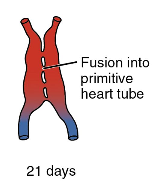
Fusion into primitive heart tube Fusion into primitive heart tube marks the stage where the paired endocardial tubes merge into a single tubular structure, a key step in heart formation. This process, guided by precise cellular and molecular interactions, enables the heart to initiate rhythmic contractions, laying the groundwork for future chamber development.
21 days 21 days represents the gestational age of the embryo, aligning with Carnegie stage 10, when the heart tube begins its functional role. At this point, the embryo measures about 2.5-3 mm, with the heart tube starting to elongate and loop, a precursor to the complex cardiac anatomy.
The Importance of Heart Tube Formation
This stage highlights the transformation of embryonic structures into a functional organ. The fusion process is a cornerstone of cardiovascular development, ensuring the embryo’s survival as it grows.
- Mechanism of Fusion: The endocardial tubes, originally separate, align and fuse along the midline due to differential growth and cellular adhesion. This fusion is regulated by genes like NKX2-5, which orchestrate cardiac cell differentiation.
- Initiation of Contractions: Following fusion, the primitive heart tube starts beating, driven by spontaneous calcium-mediated signals in the myocardial layer. This early pulsation supports the circulation of primitive blood.
- Supporting Structures: The surrounding splanchnic mesoderm contributes to the epicardium and pericardial cavity, providing a protective and nourishing environment. These layers are vital for the heart’s structural integrity.
Anatomical Features of the 21-Day Embryo
The image illustrates the heart tube’s elongated form, with color gradients indicating the direction of blood flow. This visual aid underscores the dynamic nature of early cardiac development.
By day 21, the embryo’s C-shaped curvature becomes more pronounced due to continued folding. This folding aligns the heart tube with the developing thoracic region, facilitating its integration with other systems.
- Primitive Heart Tube Structure: The tube comprises an inner endocardial layer and an outer myocardial layer, with a thin cardiac jelly layer in between. This jelly provides structural support and aids in early valve formation.
- Vascular Connections: The tube connects to the vitelline and umbilical veins, forming a rudimentary circulatory loop. These connections rely on the yolk sac and placenta for oxygenation and nutrient supply.
- Cellular Composition: The myocardial cells begin to differentiate, increasing in number and organization. This cellular maturation is essential for the heart’s contractile strength.
- Morphological Changes: The heart tube starts to bend into an S-shape, a process driven by differential growth rates. This looping establishes the future positions of the atria and ventricles.
Developmental Milestones at 21 Days
Significant advancements mark this stage, with the heart tube’s functionality marking a leap in embryonic development. The synchronization of cardiac and neural development underscores the complexity of this period.
The neural tube nears completion, while somites continue to form along the body axis. These milestones occur alongside the heart’s activation, reflecting the integrated nature of embryogenesis.
- Heart Looping: The S-shaped loop begins, guided by left-right asymmetry signals like Nodal and Pitx2. This looping is critical for proper chamber alignment and septation.
- Genetic Control: Transcription factors such as GATA4 and MEF2C enhance myocardial differentiation. Mutations in these genes can lead to defects like hypoplastic left heart syndrome.
- Blood Cell Production: Primitive erythrocytes, formed in blood islands, enter circulation via the heart tube. This early hematopoiesis supports the embryo’s metabolic demands.
- Environmental Influence: Adequate maternal nutrition, including folate, supports neural and cardiac development. Deficiencies can disrupt these critical processes.
Physiological Role of the Primitive Heart Tube
The primitive heart tube’s physiology centers on establishing circulation to meet the embryo’s growing needs. Its early beating marks the shift from passive diffusion to active blood movement.
This rudimentary pump operates under low pressure, relying on the yolk sac for oxygenation. The transition to a more complex circulatory system will refine these functions in subsequent stages.
- Contraction Dynamics: The heart tube’s peristaltic movements propel blood in a unidirectional flow. This rhythm increases in coordination as the myocardial layer thickens.
- Oxygen Transport: Primitive red blood cells carry hemoglobin with limited oxygen-binding capacity, supplemented by the yolk sac’s vitelline circulation. This system sustains the embryo until placental development.
- Pressure Gradients: The flow establishes pressure differences that guide vascular remodeling. This process ensures the heart integrates with the emerging arterial system.
- Future Adaptations: Shunts like the ductus venosus will develop to optimize fetal circulation. Understanding these adaptations aids in diagnosing congenital anomalies.
Clinical and Research Implications
Insights from the 21-day embryo inform medical practice and scientific inquiry. The heart tube’s formation is a critical window for studying congenital heart conditions.
Advances in imaging and stem cell research enhance our understanding of these early stages. These tools offer potential for both diagnosis and treatment.
- Congenital Heart Defects: Errors in heart tube looping can lead to transposition of the great arteries. Early detection via fetal echocardiography improves outcomes.
- Regenerative Potential: Stem cells can be induced to form heart tube-like structures, offering insights into cardiac repair. This approach holds promise for heart disease therapies.
- Animal Models: Chick and mouse embryos provide comparative data due to similar developmental pathways. These models help validate human embryonic studies.
- Therapeutic Innovations: Targeting growth factor pathways like VEGF may enhance heart tube development in defective embryos. Gene editing techniques are exploring this possibility.
In conclusion, this image of the 21-day embryo showcases the remarkable formation of the primitive heart tube, a vital step in cardiovascular development. This early structure sets the stage for a fully functional heart, offering valuable insights into both normal embryogenesis and potential medical interventions.

