The human heart’s development advances significantly by day 24, showcasing the emergence of key structures such as the truncus arteriosus, bulbus cordis, ventricle, atrium, and sinus venosus within the primitive heart tube. This image illustrates the heart’s looping and segmentation, a critical phase where the circulatory system begins to support the embryo’s growing demands with rhythmic contractions.
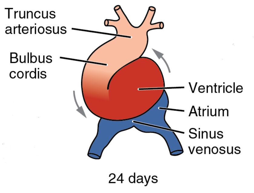
Truncus arteriosus Truncus arteriosus serves as the outflow tract, channeling blood from the heart to the developing arterial system, located at the cranial end of the heart tube. This structure will later divide into the aorta and pulmonary artery through septation, ensuring separate systemic and pulmonary circulation.
Bulbus cordis Bulbus cordis is a dilated region proximal to the truncus arteriosus, contributing to the future right ventricle and outflow tracts. Its remodeling is essential for aligning the heart’s chambers with the developing vascular network.
Ventricle Ventricle acts as the primary pumping chamber, driving blood through the bulbus cordis and truncus arteriosus, positioned centrally in the looped heart tube. This region will divide into the left and right ventricles, each specialized for its circulatory role.
Atrium Atrium receives blood returning to the heart, located caudally and beginning to form the future atrial chambers. It will expand and incorporate venous inputs, laying the groundwork for the left and right atria.
Sinus venosus Sinus venosus is the venous inflow tract, collecting deoxygenated blood from the vitelline, umbilical, and common cardinal veins at the heart’s caudal end. This structure will partially contribute to the right atrium and the sinoatrial node in later development.
24 days 24 days indicates the gestational age, corresponding to Carnegie stage 11, when the heart tube completes its looping and begins functional segmentation. The embryo, approximately 3-4 mm long, relies on this early heart activity for nutrient and oxygen distribution.
The Importance of Heart Looping and Segmentation
This stage marks a transformative period in heart development, with looping shaping the organ’s structure. The segmentation into distinct regions supports the embryo’s circulatory needs as it grows.
- Looping Process: The heart tube’s S-shaped loop forms due to differential growth rates, guided by NKX2-5 and Pitx2 signaling. This alignment positions the future atria and ventricles correctly.
- Segmentation Dynamics: The division into truncus arteriosus, bulbus cordis, ventricle, atrium, and sinus venosus reflects regional specialization. This process is driven by gene expression and cellular migration.
- Contraction Initiation: Rhythmic contractions begin, propelled by the myocardial layer’s calcium signaling. These beats establish early circulation, supporting embryonic metabolism.
Anatomical Features of the 24-Day Embryo
The image depicts the heart’s S-shaped loop, with arrows indicating blood flow direction through its segments. This visual aid highlights the dynamic nature of cardiac development at this stage.
The embryo’s continued folding positions the heart within the thoracic cavity, protecting its delicate structures. The color gradients reflect the flow from venous to arterial regions, aiding in anatomical understanding.
- Truncus Arteriosus Structure: This outflow tract connects to the aortic sac, with endothelial cells forming its inner lining. Neural crest cells will later migrate to initiate septation.
- Bulbus Cordis Anatomy: Positioned between the ventricle and truncus, this region thickens its myocardial walls. It contributes to the right ventricle and conus arteriosus.
- Ventricle Characteristics: The ventricle’s thicker walls indicate its pumping role, with trabeculae beginning to form. Its division will create the left and right ventricles.
- Atrium Details: The atrium receives blood from the sinus venosus, with early septation starting. This chamber will expand to accommodate venous return.
- Sinus Venosus Features: This venous collector merges with the atrium, with its right horn becoming prominent. It will contribute to the sinoatrial node’s pacemaker function.
Developmental Milestones at 24 Days
This phase brings significant progress, with the heart’s looping and segmentation driving circulatory advancements. The coordination with other systems underscores the complexity of embryogenesis.
The neural tube nears closure, and somites along the body axis increase, paralleling cardiac development. These milestones ensure the embryo’s holistic growth.
- Looping Completion: The S-shape loop adjusts under left-right asymmetry signals, ensuring proper chamber orientation. This process is critical for future heart function.
- Cellular Contributions: Cardiac neural crest cells migrate to the outflow tract, aiding truncus arteriosus septation. This migration prevents congenital outflow defects.
- Vascular Connections: The sinus venosus links to the vitelline and umbilical veins, forming a rudimentary circulatory loop. This network supports nutrient delivery.
- Genetic Regulation: Genes like GATA4 enhance myocardial differentiation, guiding chamber formation. Mutations can lead to ventricular septal defects.
Physiological Functions of the Looped Heart
The looped heart begins to perform essential physiological roles, marking the transition to active circulation. Its segmentation enhances blood distribution to meet embryonic needs.
The heart’s contractions strengthen, establishing a basic conduction system. This efficiency prepares the embryo for further circulatory adaptations.
- Pumping Action: The ventricle’s contractions propel blood through the bulbus cordis and truncus arteriosus. This flow supports the embryo’s metabolic demands.
- Blood Flow Patterns: Deoxygenated blood enters via the sinus venosus, while oxygenated blood exits through the truncus arteriosus. This pattern anticipates dual circulation.
- Oxygen Support: Primitive erythrocytes from blood islands carry limited oxygen, supplemented by the yolk sac. This sustains the embryo until placental circulation develops.
- Pressure Gradients: The looped structure creates pressure differences, guiding vascular remodeling. These gradients influence arterial and venous development.
Clinical and Research Implications
The 24-day embryo offers valuable insights for medical and scientific exploration. The heart’s looping and segmentation are key areas for studying congenital conditions.
Advanced imaging and stem cell research provide deeper understanding of this stage. These tools hold potential for early diagnosis and therapeutic innovations.
- Congenital Defects: Errors in looping can cause dextrocardia, detectable via ultrasound. Early intervention improves outcomes.
- Regenerative Medicine: Stem cells can be induced to form looped heart structures. This approach supports cardiac repair strategies.
- Animal Models: Chick and mouse embryos exhibit similar looping patterns. These models validate human developmental research.
- Therapeutic Targets: Modulating VEGF pathways may enhance vascular growth in defects. Gene editing explores correcting cardiac gene mutations.
In conclusion, this image of the 24-day embryo illustrates the heart’s looping and segmentation into key regions, a vital step in cardiovascular development. This early structuring supports embryonic life and provides a foundation for understanding and addressing heart-related health issues.

