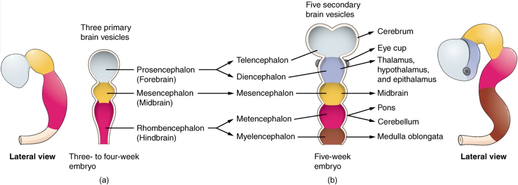
The embryonic brain undergoes remarkable transformation as it develops from the neural tube, progressing through distinct vesicle stages that shape its complex structure. This article examines a detailed image highlighting the primary vesicle stage with three regions and the secondary vesicle stage with five regions, providing insight into the early anatomical development of the brain.
Neural tube The neural tube forms the foundation of the embryonic brain and spinal cord, originating from the ectoderm layer. It undergoes folding and closure to create the initial structure for brain development.
Primary vesicle stage The primary vesicle stage marks the early division of the neural tube into three regions. This stage lays the groundwork for the brain’s major subdivisions during embryogenesis.
Forebrain (prosencephalon) The forebrain, or prosencephalon, is one of the three primary vesicles, giving rise to the telencephalon and diencephalon. It will eventually develop into structures like the cerebral hemispheres and thalamus.
Midbrain (mesencephalon) The midbrain, or mesencephalon, is another primary vesicle, connecting the forebrain and hindbrain. It evolves into regions responsible for motor control and auditory processing.
Hindbrain (rhombencephalon) The hindbrain, or rhombencephalon, forms the third primary vesicle, developing into the metencephalon and myelencephalon. It will contribute to the cerebellum, pons, and medulla oblongata.
Secondary vesicle stage The secondary vesicle stage refines the primary vesicles into five distinct regions as the brain becomes more complex. This stage reflects further specialization of brain structures.
Telencephalon The telencephalon, derived from the forebrain, develops into the cerebral hemispheres and cortex. It is responsible for higher cognitive functions like memory and reasoning.
Diencephalon The diencephalon, also from the forebrain, forms the thalamus, hypothalamus, and epithalamus. It serves as a relay station for sensory and motor signals.
Mesencephalon The mesencephalon, or midbrain, retains its name in the secondary stage, coordinating movement and processing auditory and visual reflexes. It connects the forebrain and hindbrain.
Metencephalon The metencephalon, part of the hindbrain, develops into the pons and cerebellum. It plays a key role in motor coordination and balance.
Myelencephalon The myelencephalon, the lowest hindbrain region, evolves into the medulla oblongata. It regulates vital functions such as heart rate and respiration.
Overview of Embryonic Brain Development
The brain’s development begins with the neural tube, progressing through vesicle stages. This process establishes the foundation for the central nervous system.
- The neural tube forms early in embryogenesis, closing to create a hollow structure.
- The primary vesicle stage divides the tube into three regions: forebrain, midbrain, and hindbrain.
- The secondary vesicle stage further subdivides these into five regions for specialization.
- This staged development supports the brain’s complex functionality.
- The process is critical for forming the adult brain structure.
Primary Vesicle Stage: Initial Brain Division
The primary vesicle stage initiates the brain’s regional organization. This early division sets the stage for further complexity.
- The forebrain begins as the prosencephalon, destined for higher brain functions.
- The midbrain, or mesencephalon, links forebrain and hindbrain development.
- The hindbrain, or rhombencephalon, lays the groundwork for brainstem structures.
- These regions expand and differentiate over time.
- This stage is essential for the brain’s foundational architecture.
Secondary Vesicle Stage: Enhanced Specialization
The secondary vesicle stage refines the brain into five distinct regions. This progression enhances functional diversity.
- The telencephalon develops into the cerebral cortex, supporting cognition.
- The diencephalon forms relay centers like the thalamus.
- The mesencephalon maintains its role in motor and sensory integration.
- The metencephalon contributes to the cerebellum for coordination.
- The myelencephalon becomes the medulla, regulating vital functions.
Clinical Relevance and Developmental Insights
Understanding vesicle stages aids in studying brain development and related anomalies. This knowledge is valuable for clinical applications.
- Abnormalities in the neural tube can lead to neural tube defects like spina bifida.
- Forebrain malformations may result in holoprosencephaly, affecting facial development.
- Midbrain issues can impair motor control or reflex responses.
- Hindbrain defects may cause conditions like Chiari malformation.
- Imaging techniques track these stages for diagnostic purposes.
The embryonic brain’s evolution from the neural tube through the primary and secondary vesicle stages forms a sophisticated structure that underpins all neurological functions. The progression from three to five regions highlights the increasing complexity, providing a foundation for understanding brain anatomy and addressing developmental disorders effectively.

