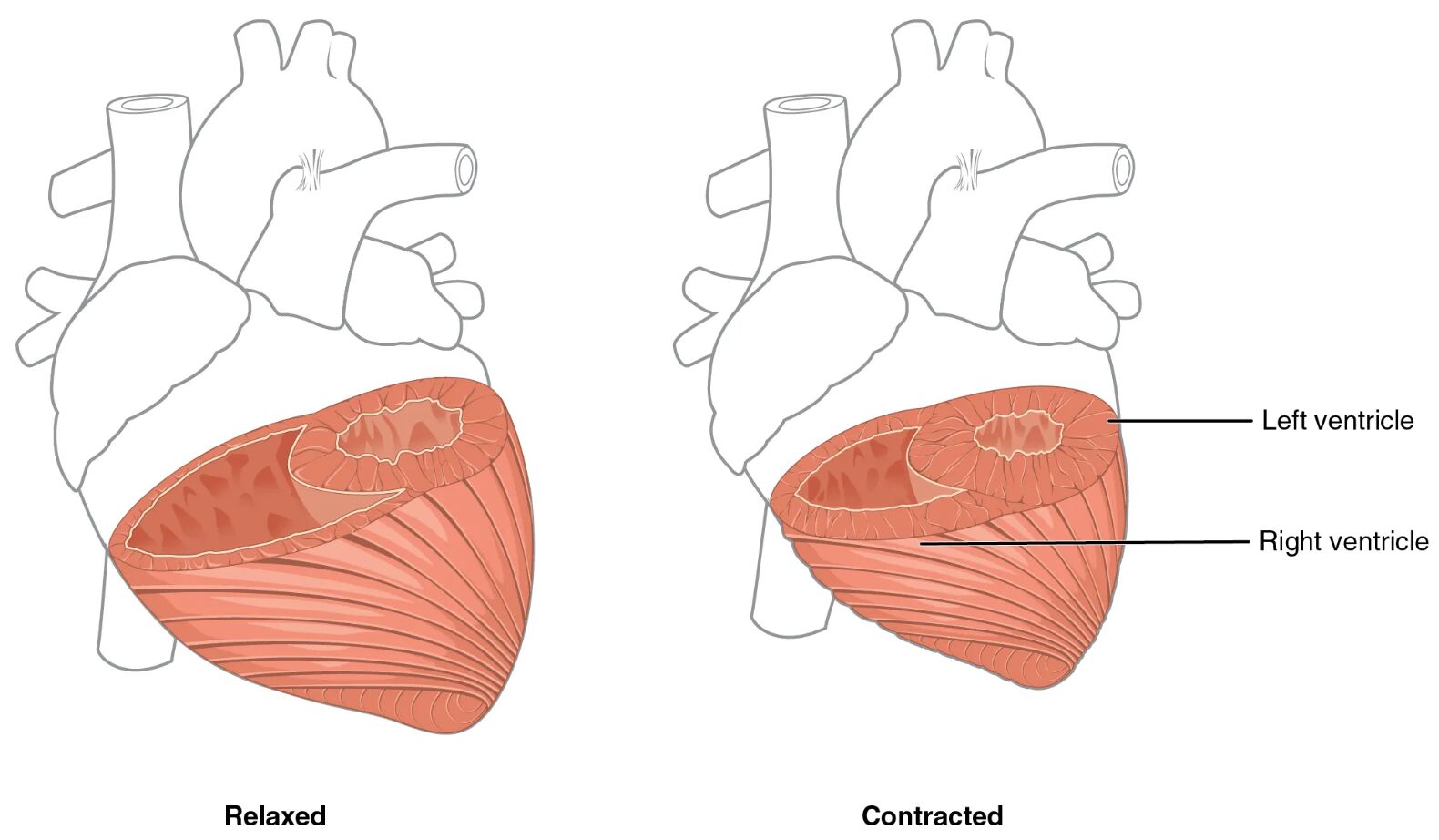
The heart’s ventricles exhibit remarkable differences in muscle thickness, reflecting their distinct roles in circulation. This diagram illustrates the left ventricle and right ventricle in both relaxed and contracting states, highlighting how the thicker myocardium of the left ventricle generates greater pressure for systemic circulation. Exploring this image provides a clear understanding of how ventricular anatomy supports the body’s dual circulatory demands.
Labelled Parts Explanation
- Left ventricle (relaxed) The left ventricle (relaxed) shows its thicker myocardium at rest, preparing to receive oxygenated blood from the left atrium. Its larger muscle mass allows it to withstand the high pressure needed for systemic circulation.
- Left ventricle (contracted) The left ventricle (contracted) demonstrates a reduced lumen as its thick myocardium contracts, ejecting blood into the aorta. This state reflects the high-pressure environment of systemic circulation.
- Right ventricle (relaxed) The right ventricle (relaxed) features a thinner myocardium, receiving deoxygenated blood from the right atrium during diastole. Its design suits the lower pressure of pulmonary circulation.
- Right ventricle (contracted) The right ventricle (contracted) shows a smaller lumen as its thinner myocardium contracts, pumping blood into the pulmonary artery. This action supports efficient gas exchange in the lungs.
- Myocardium The myocardium is the muscular layer of the heart wall, with the left ventricle’s version being significantly thicker than the right’s. This difference enables the left ventricle to generate the force required to overcome systemic resistance.
Anatomical Overview of Ventricular Muscle Thickness
The ventricles’ muscle thickness is a key adaptation to their circulatory roles. This diagram highlights the structural differences that ensure effective blood flow.
- The left ventricle (relaxed) and left ventricle (contracted) show a robust myocardium, tailored for systemic pressure.
- The right ventricle (relaxed) and right ventricle (contracted) have a thinner myocardium, optimized for pulmonary circulation.
- The lumen size varies with contraction, reflecting the ventricles’ pumping efficiency.
- Both ventricles handle the same blood volume, but their muscle mass differs to meet pressure demands.
This anatomical variation underscores the heart’s ability to balance dual circulation.
Role of the Left Ventricle in Systemic Circulation
The left ventricle’s thick musculature is designed for high-pressure output. Its structure supports the body’s systemic needs.
- The left ventricle (relaxed) fills with oxygenated blood, stretching its thick myocardium to prepare for contraction.
- The left ventricle (contracted) ejects blood into the aorta, overcoming resistance in the systemic arteries.
- The myocardium’s greater thickness provides the force needed for this high-pressure environment.
- This adaptation ensures oxygen-rich blood reaches all body tissues.
The left ventricle’s design is critical for maintaining systemic perfusion.
Function of the Right Ventricle in Pulmonary Circulation
The right ventricle’s thinner musculature suits its lower-pressure role. This structure supports pulmonary gas exchange.
- The right ventricle (relaxed) receives deoxygenated blood, with its thinner myocardium accommodating lower pressure.
- The right ventricle (contracted) pumps blood into the pulmonary artery, requiring less force than the left side.
- The myocardium’s reduced thickness reflects the lower resistance of the pulmonary circuit.
- This efficiency aids in oxygenating blood for return to the left heart.
The right ventricle’s anatomy is tailored to pulmonary demands.
Impact of Contraction on Ventricular Lumen
The ventricles’ lumen size changes with contraction, reflecting muscle action. This dynamic is vital for circulation.
- The left ventricle (contracted) shows a smaller lumen, maximizing blood ejection into the aorta.
- The right ventricle (contracted) also reduces its lumen, but to a lesser extent due to lower pressure needs.
- The left ventricle (relaxed) and right ventricle (relaxed) expand to accommodate blood volume during diastole.
- This rhythmic change ensures efficient filling and emptying of the ventricles.
The lumen’s adaptability supports the heart’s pumping cycle.
Physiological Significance of Myocardial Thickness
The myocardium’s thickness has profound physiological implications. This variation optimizes cardiac performance.
- The thicker myocardium in the left ventricle generates the pressure to overcome systemic vascular resistance.
- The thinner myocardium in the right ventricle matches the lower resistance of the pulmonary vasculature.
- This difference allows both ventricles to pump equal volumes despite varying workloads.
- The myocardium’s fiber arrangement enhances contraction efficiency across both ventricles.
This balance is essential for maintaining circulatory homeostasis.
Clinical Relevance of Ventricular Differences
Understanding ventricular muscle thickness aids in diagnosing cardiac conditions. These differences are key clinical indicators.
- Hypertrophy of the left ventricle’s myocardium can signal hypertension or aortic stenosis.
- Thinning of the right ventricle’s myocardium may indicate right heart failure or pulmonary hypertension.
- The left ventricle (contracted)’s reduced lumen can be assessed for ejection fraction in heart disease.
- Imaging like echocardiography evaluates these differences for timely intervention.
This knowledge supports effective management of cardiovascular health.
Conclusion
The differences in ventricular muscle thickness diagram offers a detailed view of how the left ventricle and right ventricle are anatomically adapted for their roles. By exploring the thicker myocardium of the left ventricle and the thinner myocardium of the right ventricle, one gains insight into the heart’s ability to handle dual circulation with equal blood volume. This understanding provides a solid basis for studying cardiovascular physiology and addressing related health issues, encouraging further exploration of the heart’s remarkable design and functional efficiency.

