The skin, as the body’s largest organ, serves as a sensory interface, with specific regions linked to the spinal nerves that transmit tactile and pain signals to the brain. This diagram illustrates dermatomes, the topographic areas of the skin corresponding to the sensory innervation of individual spinal nerves, providing a clear visual guide to this anatomical relationship. Exploring these dermatomes offers a deeper understanding of how sensory information is organized and how it can be assessed for neurological health, making it a valuable resource for anyone interested in human anatomy and physiology.
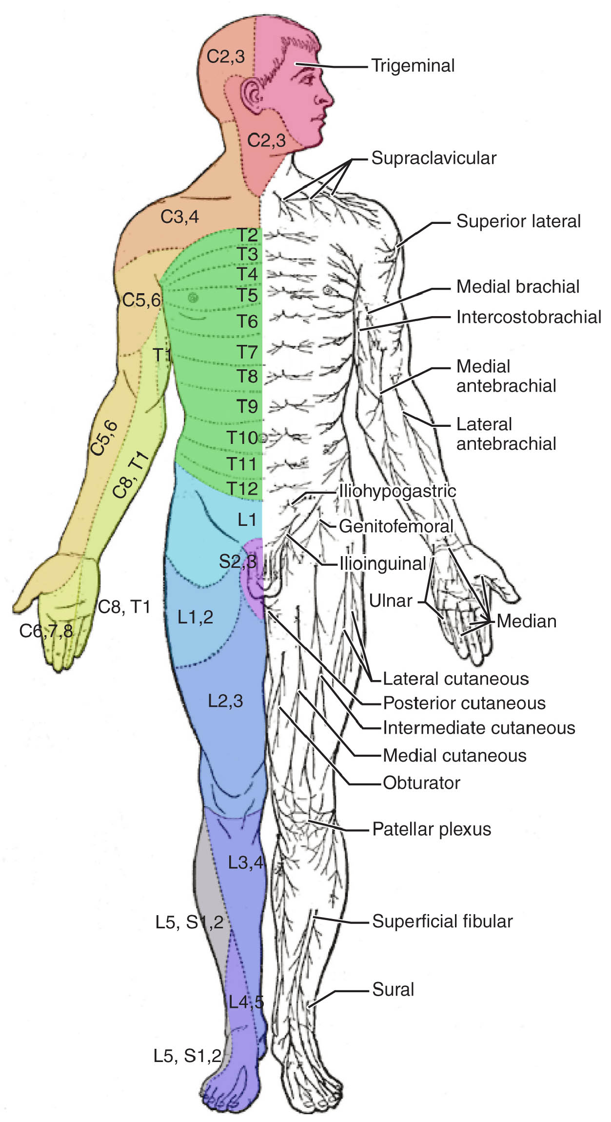
C2 The C2 dermatome covers the occipital region of the scalp and upper neck, receiving sensory input from the second cervical nerve. It plays a key role in sensing touch and pain in the back of the head, often assessed for nerve-related headaches.
C3 The C3 dermatome extends over the lateral neck and upper shoulder, innervated by the third cervical nerve. It contributes to sensation around the upper trapezius, helping detect discomfort or injury in this area.
C4 The C4 dermatome includes the lower neck and upper shoulder region, supplied by the fourth cervical nerve. It is vital for sensing pressure or pain along the clavicle and upper chest, aiding in posture-related evaluations.
T2 The T2 dermatome spans the upper chest and inner arm, linked to the second thoracic nerve. It relays sensory data from the axillary region, useful in diagnosing upper thoracic nerve issues.
T4 The T4 dermatome covers the mid-chest area, corresponding to the fourth thoracic nerve. It provides sensation over the nipple line, serving as a landmark for assessing thoracic spinal health.
T10 The T10 dermatome includes the area around the umbilicus, innervated by the tenth thoracic nerve. It marks the midline of the abdomen, often checked for abdominal pain or nerve damage.
L1 The L1 dermatome encompasses the upper groin and lower abdomen, supplied by the first lumbar nerve. It detects sensation in the inguinal region, aiding in the evaluation of lower back or pelvic issues.
L4 The L4 dermatome covers the anterior thigh and medial lower leg, linked to the fourth lumbar nerve. It is crucial for sensing touch along the shin, often tested in cases of sciatica or lumbar radiculopathy.
L5 The L5 dermatome includes the lateral leg, dorsum of the foot, and first toe, innervated by the fifth lumbar nerve. It plays a significant role in detecting foot movement and pain, commonly assessed in lower back conditions.
S1 The S1 dermatome spans the posterior thigh, lateral foot, and fifth toe, supplied by the first sacral nerve. It is essential for sensing the sole and heel, often evaluated for sciatic nerve dysfunction.
S2 The S2 dermatome covers the posterior thigh and perineal region, linked to the second sacral nerve. It contributes to sensation in the buttocks and genitals, important for diagnosing sacral nerve injuries.
S3 The S3 dermatome includes the perineal area and inner thighs, innervated by the third sacral nerve. It supports sensory input in the pelvic floor, often checked for bladder or bowel control issues.
S4-5 The S4-5 dermatome encompasses the perianal region and genitalia, supplied by the fourth and fifth sacral nerves. It is critical for sensing the anal sphincter and perineal skin, aiding in assessments of spinal cord injuries.
Anatomy of Dermatomes
Dermatomes represent the skin’s sensory map tied to spinal nerves. This diagram outlines their distribution across the body.
- The dermatomes are segmental areas defined by the spinal nerve roots, from cervical to sacral levels.
- Each dermatome overlaps slightly with adjacent ones, ensuring continuous sensory coverage.
- The cervical dermatomes (C2-C4) dominate the head, neck, and upper shoulders.
- Thoracic dermatomes (T2-T10) align with the chest and abdomen in a banded pattern.
- Lumbar and sacral dermatomes (L1-S5) cover the lower body and pelvic regions.
Sensory Innervation and Spinal Nerves
Spinal nerves provide the sensory input mapped by dermatomes. Their organization supports detailed sensation.
- Each spinal nerve exits the vertebral column and branches to innervate a specific dermatome.
- The dorsal root ganglia house the cell bodies of sensory neurons feeding into these areas.
- Cervical nerves like C2 and C3 supply the head and neck with fine touch receptors.
- Thoracic nerves, such as T4 and T10, relay visceral and somatic sensations.
- Sacral nerves (S2-S5) connect to the pelvic floor, integrating with autonomic functions.
Clinical Relevance of Dermatome Mapping
Dermatome diagrams assist in diagnosing neurological conditions. This visual tool enhances clinical accuracy.
- Loss of sensation in the C4 dermatome may indicate cervical spine injury or nerve compression.
- T10 numbness could suggest thoracic disc herniation or radiculopathy.
- L5 sensory deficits are common in herniated lumbar discs, affecting the leg and foot.
- S1 abnormalities might point to sciatica or sacral plexus damage.
- Pinprick or light touch tests map these changes to pinpoint nerve root issues.
Assessing Dermatome Function
Testing dermatomes is a practical approach to evaluate nerve health. This process reveals sensory integrity.
- Clinicians use a pinwheel or cotton swab to test sensation across dermatomes.
- The L4 dermatome is checked along the shin for reflexes like the patellar response.
- S1 sensation on the lateral foot correlates with the Achilles reflex.
- Asymmetry between sides indicates possible nerve root irritation.
- Repeated assessments track recovery or progression of nerve damage.
Neurotransmitters and Hormonal Influences
Chemical signals enhance dermatome function within the nervous system. These factors modulate sensory input.
- Glutamate excites sensory neurons, amplifying signals from dermatomes.
- Substance P mediates pain perception in areas like the S2 dermatome.
- Serotonin modulates pain thresholds across spinal nerve distributions.
- Thyroid hormones like T3 and T4 support metabolic health of sensory nerves.
- Imbalances can lead to heightened or diminished sensation in dermatomes.
Developmental and Anatomical Variations
Dermatome patterns develop early and vary slightly among individuals. This diagram reflects a standard layout.
- Embryonic segmentation establishes dermatomes during neural tube formation.
- The T10 dermatome’s umbilical alignment is a consistent developmental marker.
- Variations in overlap occur, especially in the lumbar and sacral regions.
- Aging may alter dermatome sensitivity due to nerve degeneration.
- Genetic factors influence the precise boundaries of each dermatome.
Advances in Dermatome Research
Ongoing studies refine our understanding of dermatomes. This diagram supports innovative explorations.
- MRI and nerve conduction studies map dermatome innervation with precision.
- Regenerative therapies target damaged spinal nerves affecting dermatomes.
- Virtual reality aids in simulating dermatome testing for training.
- Research into peripheral neuropathy explores dermatome-specific changes.
- These advancements enhance diagnostic and therapeutic approaches.
In conclusion, this diagram of dermatomes provides a comprehensive map of the skin’s sensory regions, linking each area to its corresponding spinal nerve for a detailed understanding of neural innervation. From the cervical dermatomes covering the head and neck to the sacral dermatomes encompassing the pelvic floor, this anatomical guide is essential for assessing sensory function and diagnosing nerve-related conditions. By leveraging this knowledge, we can improve clinical evaluations and contribute to advancements in neurological care, ensuring better outcomes for those with sensory impairments.

