Explore the vital network of blood vessels that nourish the heart muscle itself, meticulously labeled in this detailed diagram of the coronary circulation. This comprehensive overview highlights the arterial pathways that ensure the myocardium receives a continuous supply of oxygen and nutrients, essential for its tireless pumping action. Understanding coronary arteries is fundamental to comprehending heart function and the devastating impact of coronary artery disease.
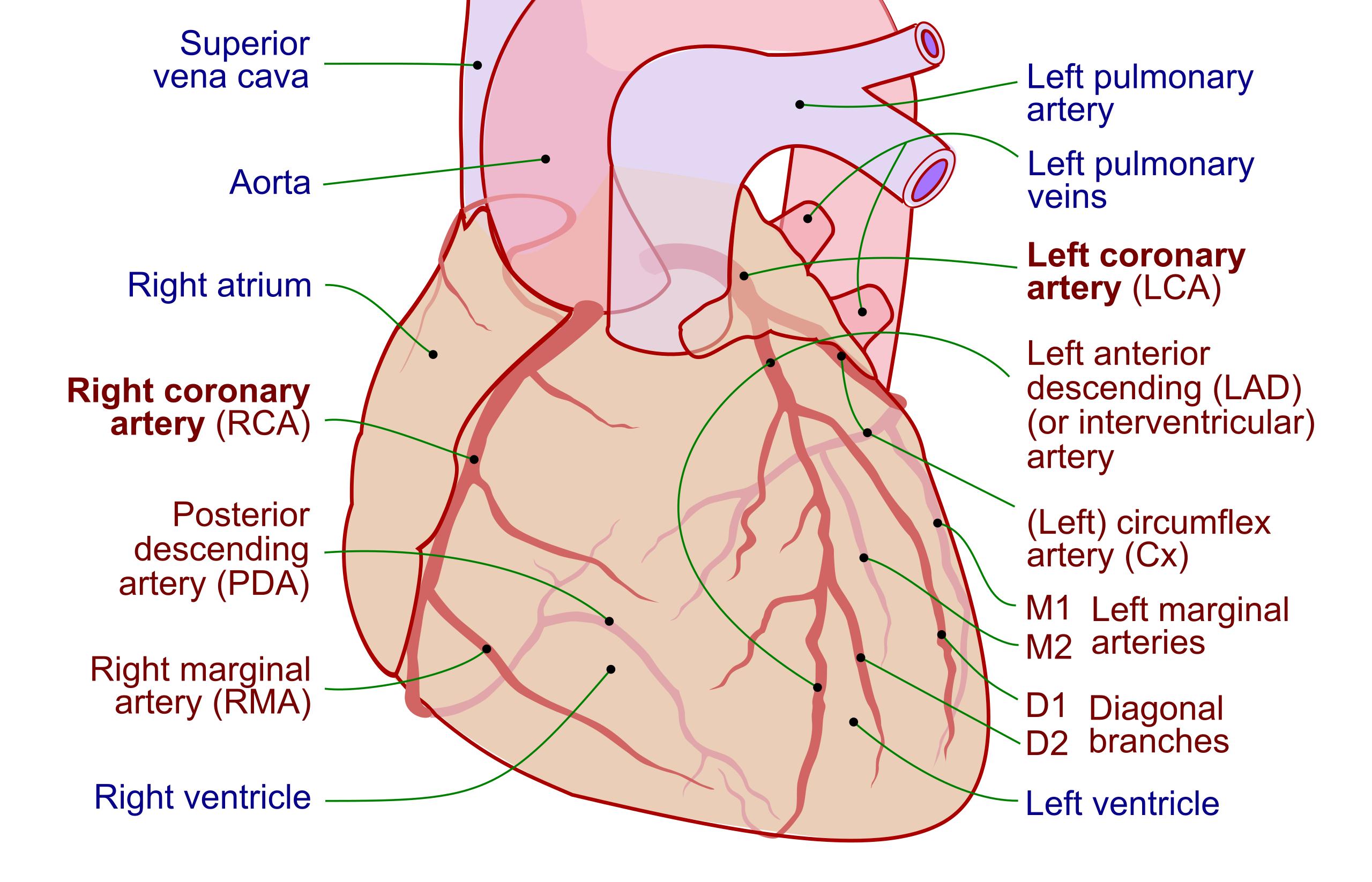
This image provides a comprehensive overview of the coronary circulation, with specific labels for the coronary arteries in red text and other key cardiac landmarks in blue text.
Superior vena cava (Landmark): This large vein collects deoxygenated blood from the upper half of the body and delivers it to the right atrium. It is a major vessel for systemic venous return, located superior to the heart.
Aorta (Landmark): The largest artery in the body, originating from the left ventricle. It is the main vessel that distributes oxygenated blood to the entire systemic circulation and is the source of the coronary arteries.
Right atrium (Landmark): The upper-right chamber of the heart that receives deoxygenated blood from the body via the superior and inferior vena cava. It acts as a collecting chamber before pumping blood into the right ventricle.
Left pulmonary artery (Landmark): This artery carries deoxygenated blood from the right ventricle to the left lung for oxygenation. It is a critical component of the pulmonary circulation.
Left pulmonary veins (Landmark): These veins return oxygenated blood from the left lung to the left atrium. They complete the pulmonary circuit, bringing freshly oxygenated blood back to the heart.
Right ventricle (Landmark): The lower-right chamber of the heart responsible for pumping deoxygenated blood to the lungs. Its muscular contractions drive blood through the pulmonary artery.
Left ventricle (Landmark): The lower-left and most muscular chamber of the heart, responsible for pumping oxygenated blood into the aorta and to the rest of the body. Its powerful contractions generate systemic blood pressure.
Left coronary artery (LCA) (Artery): This is one of the two main coronary arteries, originating from the aorta. It typically branches into the Left Anterior Descending and Left Circumflex arteries, supplying a significant portion of the left ventricle, left atrium, and interventricular septum.
Left anterior descending (LAD) (or interventricular) artery (Artery): A major branch of the LCA, often referred to as the “widowmaker” due to the high mortality associated with its blockage. It runs down the anterior surface of the heart, supplying the anterior two-thirds of the interventricular septum and parts of both ventricles.
Left circumflex artery (Cx) (Artery): Another major branch of the LCA, which wraps around the left side of the heart in the coronary sulcus. It supplies blood to the lateral wall of the left ventricle and the left atrium.
M1 Left marginal M2 arteries (Artery): These are branches off the circumflex artery, supplying the lateral wall of the left ventricle. They are important for providing blood to specific regions of the left ventricular free wall.
D1 Diagonal D2 branches (Artery): These are branches that typically arise from the LAD artery, extending diagonally over the left ventricle. They provide additional blood supply to the anterior and lateral walls of the left ventricle.
Right coronary artery (RCA) (Artery): The other main coronary artery, originating from the aorta and typically running in the coronary sulcus on the right side of the heart. It supplies blood to the right atrium, right ventricle, and often the posterior portion of the interventricular septum.
Posterior descending artery (PDA) (Artery): This artery typically arises from the RCA (in about 85% of individuals, defining “right dominance”) and runs in the posterior interventricular sulcus. It supplies the posterior one-third of the interventricular septum and parts of the posterior walls of both ventricles.
Right marginal artery (RMA) (Artery): A significant branch of the RCA, supplying the inferior and lateral walls of the right ventricle. It plays a role in vascularizing the right ventricular free wall.
The heart, a relentless pump, requires its own dedicated and robust blood supply to sustain its continuous activity. This intricate system is known as the coronary circulation, and the image meticulously details the arterial network responsible for nourishing the myocardium. Unlike other organs, the heart receives its primary blood supply during diastole, or relaxation, when the aortic valve is closed and blood can flow into the coronary arteries. Any compromise to these vital vessels can have severe consequences for cardiac health and overall bodily function.
The two main coronary arteries, the Left Coronary Artery (LCA) and the Right Coronary Artery (RCA), originate directly from the aorta and branch out to envelop the entire surface of the heart. The LCA quickly bifurcates into the Left Anterior Descending (LAD) artery, which is critical for the anterior heart wall and septum, and the Left Circumflex artery (Cx), supplying the left lateral wall. The RCA typically extends along the right side, giving rise to the Right Marginal Artery (RMA) and often the Posterior Descending Artery (PDA), which supplies the posterior aspect of the heart.
Understanding the specific territories supplied by each coronary artery is paramount in clinical practice. Blockages in these arteries, typically due to atherosclerosis (the buildup of plaque), lead to conditions like myocardial ischemia (reduced blood flow) or myocardial infarction (heart attack). For example, a blockage in the LAD artery often results in an extensive anterior wall myocardial infarction due to the large area of muscle it supplies. Therefore, precise knowledge of these arterial pathways is essential for accurate diagnosis and effective intervention in coronary artery disease.
- Coronary artery disease (CAD) is the most common type of heart disease.
- Angina pectoris, or chest pain, is a common symptom of myocardial ischemia.
- Coronary angiography is a diagnostic procedure used to visualize the coronary arteries and detect blockages.
- Coronary artery bypass grafting (CABG) and percutaneous coronary intervention (PCI) are common revascularization procedures.
This detailed illustration of the coronary arterial network is an indispensable educational tool for medical professionals, especially those in cardiology and cardiac surgery. It underscores the critical importance of a healthy coronary circulation for sustained cardiac performance and overall cardiovascular well-being. For the general public, it highlights why maintaining a heart-healthy lifestyle is vital in preventing conditions that compromise this life-sustaining blood supply to the heart.


.jpg)