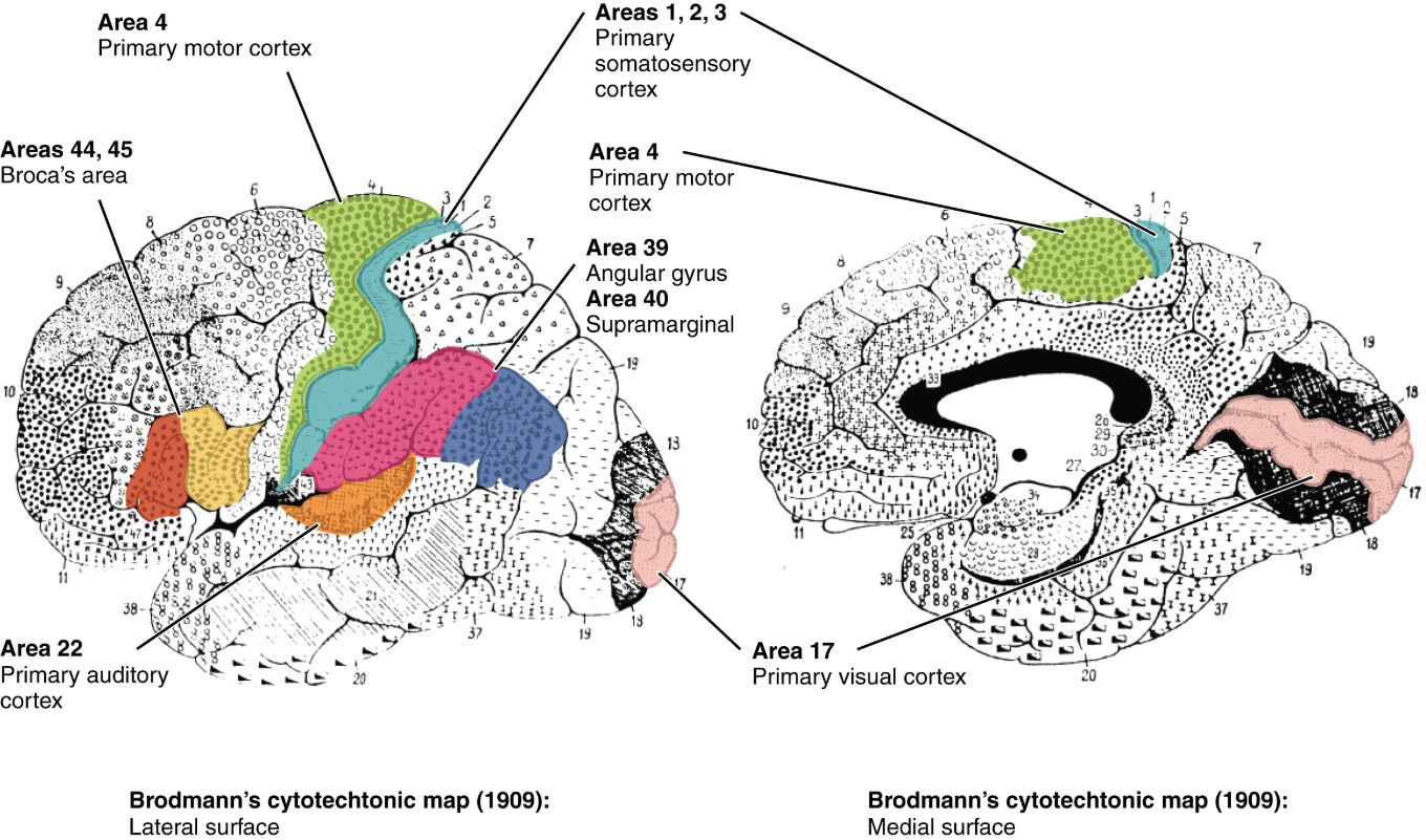
The cerebral cortex is a highly specialized region of the brain, with its functional diversity mapped by Brodmann’s areas based on microscopic cytoarchitecture. This image illustrates key areas such as Area 4, Areas 1, 2, 3, Area 39, Area 40, Areas 44, 45, and Area 22, alongside primary motor cortex, primary somatosensory cortex, angular gyrus, supramarginal gyrus, Broca’s area, primary auditory cortex, and primary visual cortex, providing a comprehensive view of cortical organization. This article explores the anatomy and significance of these regions, offering insights into their roles within the central nervous system.
Area 4
Area 4, known as the primary motor cortex, is located in the precentral gyrus and controls voluntary muscle movements. Damage to this area can lead to paralysis or impaired motor coordination on the contralateral side of the body.
Areas 1, 2, 3
Areas 1, 2, and 3 form the primary somatosensory cortex, situated in the postcentral gyrus, and process sensory input such as touch, pressure, and pain. These areas create a somatotopic map, representing different body parts with varying sensitivity.
Area 39
Area 39, the angular gyrus, is located in the parietal lobe and is involved in language processing and reading comprehension. Lesions here can result in conditions like alexia or Gerstmann syndrome, affecting spatial and mathematical skills.
Area 40
Area 40, the supramarginal gyrus, lies adjacent to the angular gyrus and contributes to language perception and spatial awareness. Dysfunction in this region may impair the ability to understand spoken language or recognize objects by touch.
Areas 44, 45
Areas 44 and 45, collectively known as Broca’s area, are located in the frontal lobe and are essential for speech production and language formulation. Damage to these areas can cause expressive aphasia, where speech output is limited despite intact comprehension.
Area 22
Area 22, part of the primary auditory cortex in the temporal lobe, processes auditory information and is crucial for sound recognition. Impairment here can lead to auditory agnosia, where sounds cannot be interpreted correctly.
Primary motor cortex
The primary motor cortex, synonymous with Area 4, initiates and fine-tunes voluntary movements through a detailed somatotopic representation. It receives input from the premotor cortex and cerebellum to ensure precise motor control.
Primary somatosensory cortex
The primary somatosensory cortex, encompassing Areas 1, 2, and 3, interprets sensory data from the body’s surface and internal structures. This region’s organization allows for the localization of tactile sensations across the body.
Angular gyrus
The angular gyrus (Area 39) integrates visual and auditory information, supporting reading and cross-modal associations. Its role extends to attention and memory processes within the parietal lobe.
Supramarginal gyrus
The supramarginal gyrus (Area 40) aids in phonological processing and sensory integration, bridging auditory and tactile inputs. It is also implicated in working memory and spatial cognition.
Broca’s area
Broca’s area (Areas 44 and 45) is critical for articulating speech and coordinating the muscles involved in talking. It works closely with Wernicke’s area to facilitate fluent language expression.
Primary auditory cortex
The primary auditory cortex (Area 22) receives sound signals via the auditory nerve and thalamus, enabling sound discrimination and localization. It is essential for processing complex auditory patterns like music or speech.
Primary visual cortex
The primary visual cortex (Area 17), located in the occipital lobe, processes visual information from the retina via the optic nerve. It is responsible for basic visual features like edges and motion, relayed to higher visual areas.
Anatomical Overview of Brodmann’s Areas
Brodmann’s areas provide a cytoarchitectural map of the cerebral cortex, dividing it into 52 regions based on cell structure. This mapping enhances our understanding of functional specialization.
- The cortex’s division into areas like 4 and 17 reflects distinct histological patterns, identified by Korbinian Brodmann in 1909.
- These areas correspond to specific functions, such as motor control in Area 4 and vision in Area 17, due to varying neuronal density and layering.
- The lateral and medial surfaces of the brain each host unique areas, contributing to a comprehensive functional map.
- Blood supply from the anterior, middle, and posterior cerebral arteries supports these regions’ high metabolic needs.
Functional Roles of Motor and Somatosensory Areas
The primary motor and somatosensory cortices are pivotal for movement and sensation. Their precise organization underpins physical interaction with the environment.
- Area 4 drives voluntary movements, with its somatotopic map controlling everything from finger dexterity to leg motion.
- Areas 1, 2, and 3 process sensory input, with greater cortical representation for areas like the hands and face due to their sensitivity.
- The motor cortex integrates signals from the basal ganglia and cerebellum, refining movement accuracy.
- Somatosensory deficits, such as numbness, can arise from lesions in these areas, necessitating targeted rehabilitation.
Language and Auditory Processing Regions
Areas involved in language and auditory processing, like Broca’s and the primary auditory cortex, are essential for communication. Their specialization supports complex cognitive tasks.
- Broca’s area (Areas 44, 45) coordinates speech muscles, with damage leading to non-fluent aphasia.
- Area 22 processes auditory signals, enabling differentiation between speech sounds and environmental noises.
- The angular and supramarginal gyri (Areas 39, 40) support language comprehension and phonological working memory.
- Auditory hallucinations in schizophrenia may involve hyperactivity in Area 22, highlighting its clinical relevance.
Visual and Higher-Order Processing Areas
The primary visual cortex and higher-order areas like the angular gyrus handle visual perception and cognitive integration. These regions are crucial for interpreting the visual world.
- Area 17 receives retinal input, processing basic visual elements before relaying to areas like 18 and 19.
- The angular gyrus integrates visual and linguistic data, aiding in reading and spatial tasks.
- Damage to Area 17 can cause cortical blindness, while higher areas may affect visual recognition.
- These regions’ connectivity with the thalamus ensures efficient sensory processing and cognitive synthesis.
Clinical Relevance and Applications
Brodmann’s areas are invaluable in diagnosing and treating neurological conditions. Their mapping guides precise medical interventions.
- Lesions in Area 4 can lead to hemiplegia, requiring physical therapy to restore function.
- Aphasia from Broca’s area damage is assessed using language tests, guiding speech therapy.
- Area 17 injuries result in visual field defects, often managed with compensatory strategies.
- Functional MRI and PET scans map these areas, aiding in epilepsy surgery or tumor resection planning.
Conclusion
Brodmann’s areas of the cerebral cortex offer a detailed blueprint of the brain’s functional landscape, with regions like Area 4, Broca’s area, and the primary visual cortex playing distinct roles. This mapping not only enhances our understanding of brain anatomy but also supports advancements in diagnosing and treating neurological disorders. Exploring these areas deepens appreciation for the cortex’s complexity and its critical role in human cognition and behavior.

