The spinal cord’s microscopic cross-section reveals a highly organized structure critical for transmitting neural impulses and coordinating reflexes. This LM × 40 micrograph of a thoracic segment, provided by the Regents of University of Michigan Medical School © 2012, displays the distinctive butterfly-shaped gray matter surrounded by white matter, highlighting horns and columns essential for sensory, motor, and autonomic functions. This detailed view offers insights into the cord’s cellular architecture and its role in central nervous system operations.
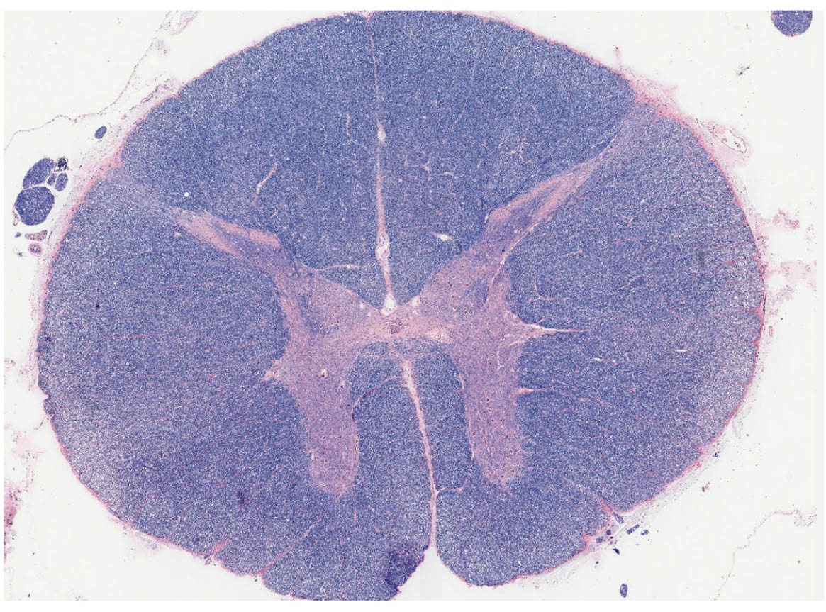
The image provided is an unlabeled microscopic view, showing the overall structure without specific annotations. Visible features include the central canal, H-shaped gray matter with posterior, lateral, and anterior horns, and surrounding white matter divided into posterior, lateral, and anterior columns.
Overview of Spinal Cord Microscopic Anatomy
The spinal cord’s histology under light microscopy distinguishes gray and white matter by staining intensity. This thoracic section exemplifies regional variations in neural tissue density.
- Gray matter stains darker due to abundant cell bodies and synapses, forming the core processing area.
- White matter appears lighter from myelin lipids, comprising axon bundles for signal conduction.
- The central canal is a small, clear lumen, often artifactually enlarged in sections.
- Thoracic specificity includes a prominent lateral horn for sympathetic neurons.
Gray Matter Horns in Detail
Gray matter horns contain neuronal somata and dendrites, facilitating local integration. Their configuration supports segmented function along the cord.
- Posterior horn processes sensory inputs, with layers of neurons classifying stimuli like touch or pain.
- Lateral horn, unique to thoracolumbar regions, houses autonomic preganglionic cells releasing acetylcholine.
- Anterior horn features large motor neurons, organized into columns innervating limb and trunk muscles.
- Intermediate zone interneurons connect horns, enabling crossed reflexes.
White Matter Columns and Tracts
White matter columns house ascending and descending fiber tracts, myelinated for rapid transmission. Their division ensures pathway segregation.
- Posterior columns carry discriminative touch and proprioception via gracile and cuneate fasciculi.
- Lateral columns include spinothalamic tracts for pain and temperature, plus corticospinal for motor control.
- Anterior columns encompass vestibulospinal tracts for balance and some uncrossed motor fibers.
- Commissural fibers link sides, allowing bilateral coordination.
Central Canal and Supporting Structures
The central canal provides a conduit for cerebrospinal fluid within the cord. Its microscopic appearance reflects ependymal lining.
- Ependyma cells facilitate fluid movement and barrier functions.
- Surrounding ventriculus terminalis in lower cord serves similar roles.
- Glial processes extend from the canal, supporting nearby neurons.
- In microscopy, the canal may contain artifacts from fixation.
Histological Staining and Cellular Components
H&E staining in this micrograph highlights nuclear basophilia in gray matter. It reveals cellular details at 40x magnification.
- Neurons show prominent nucleoli, with multipolar shapes in horns.
- Glia, including astrocytes, provide structural support and metabolic aid.
- Myelin sheaths in white matter stain pale, contrasting axonal cores.
- Blood vessels appear as clear spaces, indicating perivascular cuffs.
Physiological Roles and Integration
The spinal cord’s structure enables autonomous and relayed functions. Microscopic views correlate form with physiology.
- Sensory afferents synapse in posterior horns, ascending via white matter.
- Motor efferents from anterior horns form ventral roots for muscle activation.
- Autonomic pathways in lateral horns regulate visceral responses.
- Reflex arcs loop within gray matter, bypassing brain for speed.
Conclusion
This microscopic cross-section of the spinal cord captures the intricate balance of gray and white matter, central to neural connectivity. From sensory processing in the posterior horn to motor output in the anterior, it illustrates the cord’s foundational role in bodily control. Such views not only aid anatomical study but also inform diagnostics in spinal pathologies.

