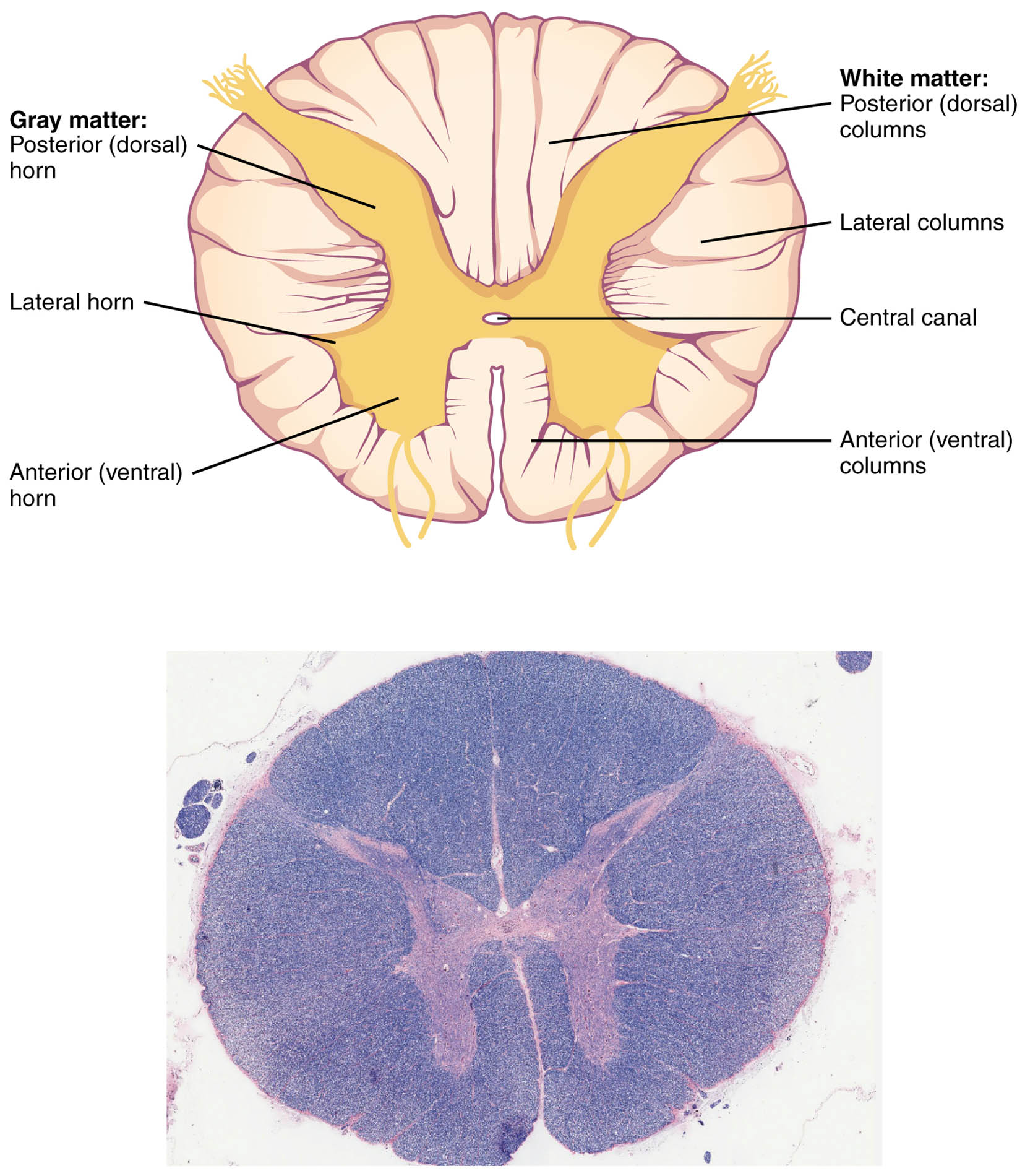
The spinal cord serves as a crucial conduit for neural signals between the brain and the body, with its cross-sectional anatomy revealing distinct gray and white matter regions essential for sensory and motor functions. This image of a thoracic spinal cord segment, accompanied by a microscopic view, illustrates the posterior (dorsal) horn, lateral horn, anterior (ventral) horn, white matter: posterior (dorsal) columns, lateral columns, anterior (ventral) columns, and central canal, providing a detailed look at its structural organization. Understanding these components enhances comprehension of neural pathways and reflex arcs.
Gray matter: Posterior (dorsal) horn
The posterior (dorsal) horn of gray matter receives sensory inputs from peripheral nerves, processing pain, temperature, and touch sensations. It contains interneurons that relay information to other spinal regions or ascend to the brain via white matter tracts.
Lateral horn
The lateral horn, present in thoracic and upper lumbar segments, houses preganglionic sympathetic neurons that regulate autonomic functions like vasoconstriction and sweat gland activity. These neurons project to sympathetic chain ganglia, influencing visceral organs and homeostasis.
Anterior (ventral) horn
The anterior (ventral) horn contains motor neurons that innervate skeletal muscles, facilitating voluntary and reflex movements. Alpha motor neurons here form the final common pathway for muscle contraction, modulated by upper motor neuron inputs.
White matter: Posterior (dorsal) columns
The posterior (dorsal) columns of white matter transmit fine touch, vibration, and proprioceptive information ascending to the brain via the medial lemniscus pathway. These myelinated tracts, including fasciculus gracilis and cuneatus, maintain somatotopic organization for precise localization.
Lateral columns
The lateral columns encompass descending motor tracts like the lateral corticospinal and rubrospinal, controlling voluntary limb movements, as well as ascending spinothalamic tracts for pain and temperature. This region supports crossed pathways, ensuring contralateral control.
Central canal
The central canal is a cerebrospinal fluid-filled channel running the length of the spinal cord, continuous with brain ventricles. Lined by ependymal cells, it facilitates nutrient exchange and waste removal, contributing to spinal cord protection.
Anterior (ventral) columns
The anterior (ventral) columns include descending vestibulospinal and reticulospinal tracts for posture and balance, along with some ascending fibers. These tracts help stabilize the body during movement and integrate reflexive responses.
Anatomical Structure of the Spinal Cord
The spinal cord’s cross-section displays a butterfly-shaped gray matter core surrounded by white matter. This organization supports efficient signal transmission and processing.
- Gray matter consists of neuronal cell bodies, dendrites, and synapses, divided into horns for functional specialization.
- White matter comprises myelinated axons grouped into columns or funiculi, facilitating long-distance communication.
- The thoracic level features a prominent lateral horn, absent in cervical and sacral regions.
- Protective meninges—dura, arachnoid, and pia—enclose the cord, with cerebrospinal fluid cushioning impacts.
Gray Matter Components and Functions
Gray matter forms the processing hub within the spinal cord. Its horn divisions reflect sensory, motor, and autonomic roles.
- The posterior horn integrates afferent signals via Rexed laminae I-VI, with lamina II (substantia gelatinosa) modulating pain.
- Lateral horn neurons release acetylcholine at preganglionic synapses, activating postganglionic adrenergic fibers.
- Anterior horn includes lower motor neurons organized somatotopically, with medial groups innervating axial muscles.
- Interneurons in intermediate zones coordinate reflexes, such as the withdrawal reflex.
White Matter Tracts and Pathways
White matter enables bidirectional neural traffic between the periphery and brain. Its columnar arrangement preserves topographic mapping.
- Posterior columns ascend ipsilaterally, decussating in the medulla to form the medial lemniscus.
- Lateral columns house the anterolateral system for crude touch and nociception, crossing within one to two segments.
- Anterior columns support uncrossed pyramidal tracts in some species, but primarily extrapyramidal in humans.
- Myelination by oligodendrocytes accelerates conduction, with demyelination disrupting function.
Significance of the Central Canal
The central canal represents a vestige of embryonic neural tube development. It maintains internal environment stability.
- Ependymal cilia promote cerebrospinal fluid flow, preventing stagnation.
- In adults, the canal may obliterate caudally, but persists rostrally linking to the fourth ventricle.
- Surrounding gray commissure connects left and right horns, allowing bilateral integration.
- Pathological enlargement, or syringomyelia, can compress adjacent tissues.
Microscopic View and Histology
The microscopic image reveals cellular details under low magnification. It highlights tissue staining differences for identification.
- H&E staining shows basophilic gray matter due to Nissl substance in neurons.
- White matter appears lighter from lipid-rich myelin sheaths.
- Glial cells, including astrocytes and microglia, support neuronal health.
- Blood vessels penetrate via perivascular spaces, supplying oxygen and nutrients.
Physiological Integration in the Spinal Cord
The spinal cord coordinates reflexes and relays information seamlessly. Gray and white matter interplay ensures responsive neural networks.
- Sensory afferents enter via dorsal roots, synapsing in posterior horns before ascending.
- Motor efferents exit ventral roots, modulated by descending tracts in white matter.
- Autonomic outflows from lateral horns regulate visceral functions independently.
- Segmental organization allows localized reflexes while permitting supraspinal override.
Conclusion
This cross-section of the spinal cord, with its detailed gray and white matter divisions, illustrates the organ’s sophisticated architecture for neural communication. From the posterior (dorsal) horn processing sensations to the anterior columns conveying motor commands, each component contributes to bodily harmony. Appreciating this anatomy deepens insight into nervous system operations and informs clinical approaches to spinal disorders.

