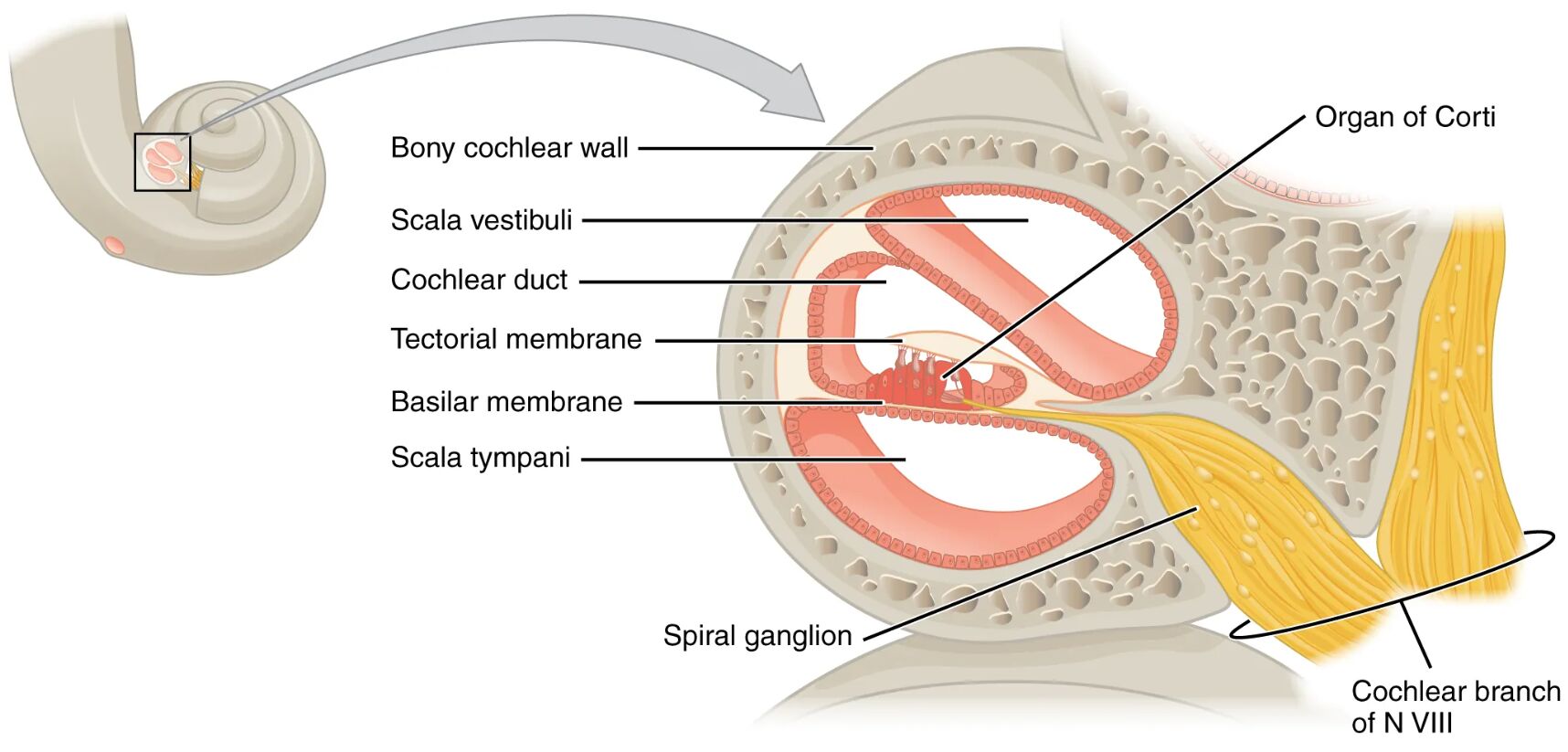
The cochlea, a spiral marvel within the inner ear, plays a pivotal role in transforming sound vibrations into electrical signals for hearing, with its internal structure revealed in this cross-sectional image. This image highlights the scala tympani, scala vestibuli, and cochlear duct, alongside the organ of Corti, which houses the hair cells essential for audition. This article provides a detailed examination of these components, offering insights into their anatomical arrangement and physiological significance in the auditory process.
Labeled Parts of the Cochlea
Scala tympani The scala tympani is a fluid-filled chamber in the lower part of the cochlea, extending from the apex to the round window, where it dissipates pressure waves. It plays a crucial role in maintaining fluid equilibrium, allowing the cochlea to process sound without damaging its delicate structures.
Scala vestibuli The scala vestibuli is an upper fluid-filled chamber that receives pressure waves from the oval window, transmitting them toward the cochlear apex. It works in tandem with the scala tympani, facilitating the movement of fluid that stimulates auditory hair cells.
Cochlear duct The cochlear duct, or scala media, is a triangular chamber located between the scala vestibuli and scala tympani, containing endolymph and the organ of Corti. It houses the sensory apparatus for hearing, separating the perilymph-filled scalae with its specialized membrane.
Organ of Corti The organ of Corti is a structure within the cochlear duct, resting on the basilar membrane, and contains mechanoreceptor hair cells that detect sound vibrations. These hair cells convert mechanical energy into electrical signals, which are transmitted to the auditory nerve.
Basilar membrane The basilar membrane supports the organ of Corti and varies in width and stiffness along the cochlea, tuning it to different sound frequencies. Its movement in response to fluid pressure waves is essential for the frequency-specific activation of hair cells.
Hair cells Hair cells are specialized mechanoreceptors within the organ of Corti, with stereocilia that bend in response to sound-induced fluid motion. They generate electrical impulses that are sent to the brain via the cochlear nerve, enabling the perception of sound.
Anatomical Overview of the Cochlea
The cochlea’s cross-sectional view reveals a sophisticated arrangement of fluid-filled chambers and sensory structures, optimized for sound processing. This internal design ensures that sound vibrations are efficiently converted into neural signals.
- Chamber division: The scala tympani and scala vestibuli flank the cochlear duct, creating a dual fluid system that supports pressure wave transmission.
- Sensory hub: The organ of Corti, nestled within the cochlear duct, is the primary site for auditory transduction, anchored by the basilar membrane.
- Fluid dynamics: Perilymph in the scalae and endolymph in the cochlear duct interact to propagate and modulate sound waves.
- Structural support: The basilar membrane’s gradient in thickness allows it to resonate at specific frequencies, enhancing pitch discrimination.
- Cellular precision: Hair cells are strategically positioned to respond to fluid movements, forming the interface between mechanical and electrical signals.
Physiological Functions of the Cochlea
The cochlea transforms mechanical sound waves into electrical impulses, enabling the perception of pitch, volume, and timbre. Its physiological processes are finely tuned to handle a wide range of auditory inputs.
- Pressure wave initiation: Vibrations from the oval window create pressure waves in the scala vestibuli, which travel through the cochlear fluid.
- Frequency analysis: The basilar membrane’s varying stiffness filters these waves, with different regions responding to specific sound frequencies.
- Hair cell activation: Hair cells within the organ of Corti bend with fluid motion, releasing neurotransmitters to activate auditory nerve fibers.
- Signal transmission: Electrical impulses generated by hair cells are relayed to the brain, where they are interpreted as sound.
- Pressure balance: The scala tympani absorbs excess pressure via the round window, maintaining cochlear stability during sound processing.
Developmental and Structural Dynamics
The cochlea develops during embryogenesis, with its chambers and sensory structures maturing to support hearing by early postnatal life. This development reflects a coordinated growth process essential for auditory function.
- Embryonic formation: The cochlear duct forms from the otic vesicle, with the scalae developing as perilymphatic spaces around it.
- Membrane development: The basilar membrane grows with a gradient of stiffness, enabling frequency-specific responses as the cochlea spirals.
- Hair cell differentiation: Hair cells emerge within the organ of Corti, with their stereocilia aligning to detect fluid motion.
- Fluid establishment: Perilymph and endolymph fill the scalae and cochlear duct, respectively, during fetal development to support sound transduction.
- Postnatal refinement: The cochlea’s sensitivity to sound improves as neural connections with hair cells strengthen after birth.
Clinical Relevance and Cochlear Health
Understanding the cochlea’s structure is vital for diagnosing and managing hearing-related conditions. Clinical assessments often target these components to identify auditory impairments.
- Sensorineural hearing loss: Damage to hair cells, often from noise exposure or ototoxic drugs, can impair sound transduction within the organ of Corti.
- Presbycusis: Age-related degeneration of the basilar membrane and hair cells leads to high-frequency hearing loss.
- Meniere’s disease: Excessive endolymph in the cochlear duct can cause pressure imbalances, resulting in vertigo and hearing issues.
- Diagnostic tools: Audiometry and cochlear imaging assess hair cell function and fluid dynamics for accurate diagnosis.
- Therapeutic options: Treatments include cochlear implants for severe hair cell loss or medications to manage fluid imbalances.
In conclusion, the cross section of the cochlea reveals a meticulously designed structure that bridges mechanical sound waves to neural perception, with the scala tympani, scala vestibuli, and organ of Corti at its core. This image underscores the cochlea’s role as the auditory powerhouse, where the basilar membrane and hair cells orchestrate the transformation of vibrations into the rich tapestry of sound we experience. Exploring these elements provides a deeper appreciation for hearing mechanisms and informs strategies to preserve auditory health.

