The coronary circulation is a crucial network that supplies oxygen and nutrients to the heart muscle, and this image presents a detailed posterior view of its prominent surface vessels. By illustrating the arteries that nourish the back of the heart, the diagram offers a comprehensive look at their anatomical distribution and significance in cardiac function. Exploring this illustration deepens understanding of the heart’s blood supply and its role in sustaining cardiovascular health.
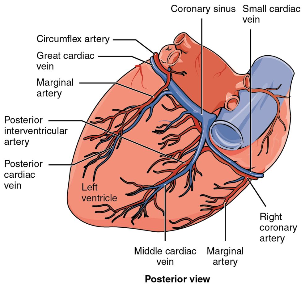
Right coronary artery: The right coronary artery originates from the aorta and supplies blood to the right atrium, right ventricle, and parts of the left ventricle via its branches. It plays a vital role in supporting the heart’s right side and maintaining electrical conduction through the atrioventricular node.
Circumflex artery: The circumflex artery, a branch of the left coronary artery, extends along the left atrioventricular groove on the posterior surface, providing blood to the lateral and posterior walls of the left ventricle. Its extensive reach ensures adequate perfusion to critical areas of the heart.
Posterior descending artery: The posterior descending artery, often a branch of the right coronary artery, runs along the posterior interventricular groove, delivering blood to the posterior walls of both ventricles and the inferior septum. It is essential for supporting the heart’s posterior region and overall stability.
Anatomical Structure of Coronary Circulation
The heart’s vascular network is meticulously designed to sustain its muscular walls, and this posterior view highlights its key arteries. The diagram provides a focused perspective on the vessels that ensure myocardial health from the back.
- The right coronary artery emerges from the aortic root, branching to cover the right heart.
- The circumflex artery curves around to the posterior, nourishing the left ventricular wall.
- The posterior descending artery targets the interventricular septum and posterior regions.
- This layout reflects the heart’s reliance on a balanced blood supply across its surfaces.
The posterior view complements the anterior, revealing the full coronary network.
Functional Roles in Cardiac Perfusion
Coronary arteries deliver oxygen-rich blood during diastole, and this image showcases their distribution on the posterior heart. Their paths support the heart’s continuous workload in this region.
- The right coronary artery feeds the right ventricle, crucial for pulmonary circulation.
- The circumflex artery ensures lateral and posterior left ventricular perfusion.
- The posterior descending artery supplies the septum, supporting coordinated contraction.
- This network maintains cardiac muscle vitality, especially during increased demand.
Blockages can lead to posterior wall infarction if untreated.
Physical Characteristics and Clinical Relevance
The physical traits of these coronary arteries reflect their adaptation to the heart’s needs. Their structure influences their vulnerability to disease and diagnostic approaches.
- The right coronary artery’s path varies, often dominating in right-dominant hearts.
- The circumflex artery’s posterior course makes it susceptible to occlusion.
- The posterior descending artery’s location in the groove aids in targeted imaging.
- Coronary angiography assesses these vessels for stenosis, guiding interventions like bypass surgery.
Their posterior position can complicate surgical access.
Importance in Cardiac Health
Preserving the patency of coronary circulation is essential for long-term heart function. The health of these arteries directly impacts cardiac endurance and recovery.
- A healthy right coronary artery prevents right-sided heart failure.
- The circumflex artery’s integrity reduces risk of posterior wall damage.
- The posterior descending artery’s flow guards against septal dysfunction.
- Lifestyle modifications, like quitting smoking, protect these vessels from atherosclerosis.
Regular stress testing monitors their condition effectively.
Conclusion
This diagram of coronary circulation from the posterior view provides a detailed illustration of the right coronary artery, circumflex artery, and posterior descending artery, showcasing their roles in heart perfusion. By highlighting the vascular network that sustains the heart’s posterior surface, it emphasizes the importance of maintaining these arteries for optimal cardiac function. This understanding fosters greater awareness of coronary anatomy and the need to safeguard heart health through proactive care.

