Delve into the intricate anatomical structure of the human heart with this detailed diagram, highlighting its chambers, valves, and major associated blood vessels. This comprehensive overview is essential for understanding how this vital organ functions as a powerful dual pump, efficiently circulating blood throughout the body. A clear grasp of these anatomical components is fundamental to comprehending cardiac physiology and identifying the origins of various cardiovascular conditions.
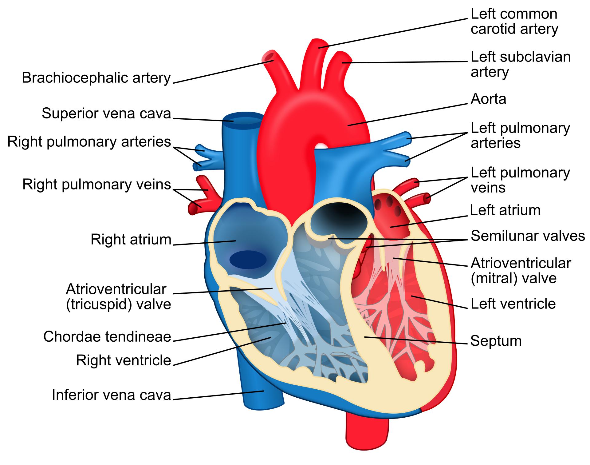
Left common carotid artery: This major artery branches off the aorta and ascends into the neck, providing oxygenated blood primarily to the left side of the head and neck. It is a critical component of the systemic circulation, ensuring blood supply to the brain and facial structures.
Left subclavian artery: Originating from the aorta, this artery supplies oxygenated blood to the left upper limb and parts of the chest and neck. Its branching pattern contributes to the extensive vascular network of the upper body.
Aorta: The aorta is the largest artery in the body, emerging from the left ventricle. It arches over the heart, giving rise to several major arteries that distribute oxygenated blood to the entire systemic circulation.
Left pulmonary arteries: These arteries carry deoxygenated blood from the right ventricle to the left lung. They are unique among arteries for transporting deoxygenated blood.
Left pulmonary veins: These veins return oxygenated blood from the left lung to the left atrium. They are essential for completing the pulmonary circuit and delivering oxygen-rich blood for systemic distribution.
Left atrium: The left atrium is the upper-left chamber of the heart that receives oxygenated blood from the lungs via the pulmonary veins. It then pumps this blood into the left ventricle.
Semilunar valves: These valves, including the aortic and pulmonary valves, are located at the exits of the ventricles, preventing the backflow of blood from the arteries into the ventricles during cardiac diastole. They are critical for maintaining unidirectional blood flow out of the heart.
Atrioventricular (mitral) valve: Also known as the bicuspid valve, this valve is situated between the left atrium and the left ventricle. It prevents the backflow of oxygenated blood into the left atrium when the left ventricle contracts.
Left ventricle: The left ventricle is the lower-left chamber of the heart and the most muscular chamber, responsible for pumping oxygenated blood into the aorta and to the rest of the body. Its powerful contractions generate the high pressure needed for systemic circulation.
Septum: The septum is the muscular wall that divides the heart into right and left sides, specifically separating the ventricles (interventricular septum) and atria (interatrial septum). It prevents the mixing of oxygenated and deoxygenated blood, ensuring efficient circulation.
Brachiocephalic artery: This is the first artery to branch off the aortic arch, supplying blood to the right arm and the right side of the head and neck. It further divides into the right common carotid artery and the right subclavian artery.
Superior vena cava: The superior vena cava is a large vein that collects deoxygenated blood from the upper half of the body (head, neck, and upper limbs) and delivers it to the right atrium. It is one of the primary vessels for systemic venous return.
Right pulmonary arteries: These arteries carry deoxygenated blood from the right ventricle to the right lung. Along with the left pulmonary arteries, they facilitate the crucial process of gas exchange in the lungs.
Right pulmonary veins: These veins return oxygenated blood from the right lung to the left atrium. They are vital for bringing freshly oxygenated blood back to the heart for systemic distribution.
Right atrium: The right atrium is the upper-right chamber of the heart, receiving deoxygenated blood from the body via the superior and inferior vena cava. It then pumps this blood into the right ventricle.
Atrioventricular (tricuspid) valve: This valve is located between the right atrium and the right ventricle. It prevents the backflow of deoxygenated blood into the right atrium when the right ventricle contracts.
Chordae tendineae: These are tough, fibrous cords that connect the cusps of the atrioventricular valves (tricuspid and mitral) to the papillary muscles within the ventricles. They prevent the valve leaflets from prolapsing into the atria during ventricular contraction.
Right ventricle: The right ventricle is the lower-right chamber of the heart, responsible for pumping deoxygenated blood to the lungs through the pulmonary arteries. Its contractions are essential for the pulmonary circulation.
Inferior vena cava: The inferior vena cava is a large vein that collects deoxygenated blood from the lower half of the body (trunk, abdomen, and lower limbs) and delivers it to the right atrium. It is another primary vessel for systemic venous return.
The human heart is an extraordinary organ, functioning as the central pump of the circulatory system. This comprehensive diagram provides an excellent overview of its intricate internal and external anatomy, showcasing the four chambers, four valves, and the major arteries and veins that connect to it. The heart operates as a double pump, with the right side receiving deoxygenated blood from the body and sending it to the lungs, while the left side receives oxygenated blood from the lungs and propels it throughout the rest of the body. This meticulous separation and coordinated action are fundamental to sustaining life.
The efficiency of blood flow is largely attributed to the precise functioning of the heart’s valves. The atrioventricular valves (tricuspid and mitral) regulate blood flow between the atria and ventricles, while the semilunar valves (aortic and pulmonary) control blood flow out of the ventricles into the great arteries. Structures like the chordae tendineae play a critical role in preventing valve prolapse, ensuring unidirectional flow. Any structural or functional abnormality in these valves can lead to various cardiovascular diseases, impacting the heart’s ability to pump effectively.
Furthermore, the diagram highlights the key great vessels that transport blood to and from the heart and body. The aorta, for instance, is the largest artery responsible for distributing oxygenated blood throughout the systemic circulation, while the superior and inferior vena cava return deoxygenated blood. Understanding these complex interconnections is vital for comprehending how the heart maintains homeostasis and responds to physiological demands. Conditions such as congenital heart defects, which can involve an abnormal septum or malformed valves, directly impact the efficiency of this complex system.
- The heart beats approximately 60-100 times per minute at rest.
- The muscular wall of the left ventricle is significantly thicker due to its role in pumping blood throughout the entire body.
- The septum’s integrity is crucial; a hole in the septum (septal defect) can lead to mixing of oxygenated and deoxygenated blood.
- The great vessels ensure that blood is efficiently delivered to and removed from all parts of the body.
This detailed anatomical depiction is an invaluable resource for medical students, healthcare professionals, and anyone seeking a deeper understanding of the human heart. A thorough knowledge of these structures and their functions is the foundation for diagnosing heart conditions, developing effective treatments, and promoting overall cardiovascular health. The heart’s intricate design is a testament to the marvel of human biology, performing its tireless work to sustain life.

