Veins and venules are essential components of the circulatory system, responsible for returning deoxygenated blood to the heart, with distinct structural differences that support their roles. This image provides a comparative view of large veins, medium-sized veins, and venules, highlighting their layered anatomy and unique features like valves that prevent backflow.
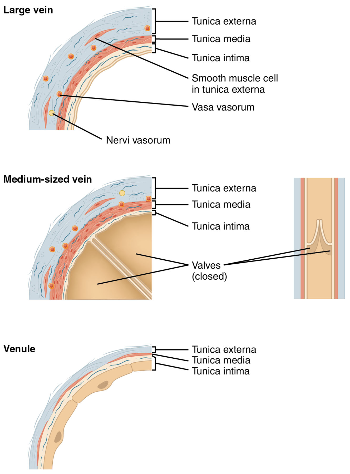
Tunica externa The tunica externa is the outermost layer of veins and venules, composed of connective tissue that provides structural support and flexibility. It anchors the vessel to surrounding tissues and contains nerves and small blood vessels, known as vasa vasorum, to nourish the wall.
Tunica media The tunica media is the middle layer, consisting of smooth muscle cells and elastic fibers that help regulate vessel diameter. In veins, this layer is thinner compared to arteries, allowing greater distensibility to accommodate varying blood volumes.
Tunica intima The tunica intima is the innermost layer, lined with endothelial cells that minimize friction and facilitate blood flow. In medium-sized veins, it includes valves that prevent backflow, a feature less prominent in venules.
Smooth muscle cell in tunica externa Smooth muscle cells in the tunica externa are sparse but contribute to the vessel’s ability to adjust its shape. They provide additional support and aid in maintaining venous tone, particularly in larger veins.
Vasa vasorum The vasa vasorum are small blood vessels within the tunica externa that supply oxygen and nutrients to the vein wall. They are more prominent in larger veins due to the thicker walls requiring nourishment.
Nervi vasorum The nervi vasorum are nerves within the tunica externa that regulate vascular tone and respond to autonomic signals. They help coordinate the contraction and relaxation of the vessel wall to manage blood flow.
Valves (closed) Valves are flap-like structures in medium-sized veins that prevent the backflow of blood, especially against gravity. When closed, they ensure unidirectional flow toward the heart, a feature absent in venules.
The Role of Veins and Venules in Circulation
Veins and venules play a critical role in returning deoxygenated blood to the heart. Their structure supports the body’s ability to maintain venous return under varying conditions.
- Tunica externa provides a protective sheath, anchoring veins to surrounding tissues.
- The thinner tunica media in veins allows them to act as blood reservoirs, holding up to 70% of the body’s blood volume.
- Valves assist in venous return, especially in the lower extremities where gravity poses a challenge.
- Venules serve as the initial collectors of blood from capillaries, transitioning it to larger veins.
Anatomical Differences Across Vein Sizes
The anatomy of large veins, medium-sized veins, and venules varies significantly based on their size and function. These differences reflect their specialized roles in the circulatory system.
- Large veins feature a well-developed tunica externa with vasa vasorum and nervi vasorum for nourishment and control.
- Medium-sized veins include valves to prevent backflow, a feature less critical in larger or smaller vessels.
- The tunica intima remains consistent but adapts with valves in medium-sized veins for efficient flow.
- Venules lack valves and have a minimal tunica media, reflecting their role as transition vessels.
Physiological Functions and Importance
The layered structure of veins and venules supports their physiological roles in blood transport. This design ensures efficient circulation and adaptation to bodily needs.
- The tunica media allows veins to expand during increased venous return, such as after exercise.
- Valves in medium-sized veins prevent pooling of blood, reducing the risk of edema.
- The tunica externa with smooth muscle cells helps maintain venous pressure during changes in posture.
- Venules facilitate the exchange of hormones like T3 and T4 from the thyroid gland as blood moves toward veins.
Clinical Significance of Veins and Venules
Understanding the anatomy of veins and venules can provide insights into circulatory health. Alterations in their structure may indicate or contribute to various conditions.
- Weak valves can lead to varicose veins, causing discomfort and swelling in the legs.
- Thickening of the tunica media in veins may occur in chronic venous insufficiency.
- Damage to the tunica intima can increase the risk of thrombosis in larger veins.
- Venule dysfunction may affect capillary exchange, impacting tissue nutrition.
Comparison with Arteries and Capillaries
Veins and venules differ from arteries and capillaries due to their specific roles in the circulatory system. This comparison highlights their unique adaptations.
- Unlike arteries, veins have a thinner tunica media and rely on valves for flow direction.
- Capillaries lack the layered structure of veins, focusing solely on exchange.
- The tunica externa is more prominent in veins than in arteries, providing flexibility.
- Venules transition from the thin-walled capillaries to the more robust vein structure.
Maintenance and Regulation of Veins and Venules
The body regulates veins and venules to ensure efficient blood return. These mechanisms adapt to physiological demands and maintain circulatory integrity.
- The nervi vasorum respond to sympathetic signals, adjusting venous tone.
- Smooth muscle cells in the tunica externa contract to aid venous return during muscle movement.
- The vasa vasorum ensure the tunica externa remains nourished, supporting vessel health.
- Valves open and close dynamically with blood pressure changes to prevent reflux.
In conclusion, the comparative anatomy of large veins, medium-sized veins, and venules, as shown in this image, illustrates the circulatory system’s adaptability. With layers like the tunica externa, tunica media, and tunica intima, along with features like valves, vasa vasorum, nervi vasorum, and smooth muscle cells, these vessels ensure efficient blood return. Exploring these structures enhances our understanding of how the venous system supports overall health and responds to physiological challenges.

