The nerve trunk, a vital component of the peripheral nervous system, reveals its intricate layers when viewed under a microscope, showcasing the protective and supportive roles of connective tissue. This high-magnification image highlights the endoneurium, perineurium, and epineurium, offering a detailed look at how these structures safeguard nerve fibers and facilitate signal transmission. Exploring this microscopic anatomy provides a deeper understanding of nerve function and its importance in maintaining bodily coordination.
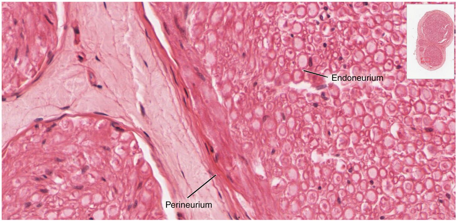
Labeled Structures in Nerve Trunk
This section provides a detailed explanation of each labeled component in the microscopic view, emphasizing their anatomical and functional significance.
Endoneurium The endoneurium is a delicate layer of connective tissue surrounding individual nerve fibers within a fascicle, providing insulation and structural support. It houses Schwann cells that produce myelin sheaths, enhancing the speed and efficiency of nerve impulse conduction.
Perineurium The perineurium is a multilayered sheath of connective tissue encasing each fascicle, forming a protective barrier that maintains a stable internal environment. It contributes to the blood-nerve barrier, regulating the exchange of substances to shield axons from potential damage.
Epineurium The epineurium is the outermost connective tissue layer enveloping the entire nerve trunk, offering mechanical protection against external forces and stretching. It contains blood vessels and adipose tissue, ensuring nutrient supply and cushioning to support nerve health.
Anatomy of the Nerve Trunk
The nerve trunk’s layered structure is a testament to its role in protecting and organizing nerve fibers. This anatomical design ensures both durability and functionality in signal transmission.
- The endoneurium, a fine network of collagen fibers, surrounds each axon, providing a microenvironment that supports myelination and nutrient delivery.
- The perineurium, with its tight junctions, creates a diffusion barrier, maintaining an optimal ionic balance for axonal function within fascicles.
- The epineurium, a dense collagenous layer, bundles multiple fascicles into a cohesive nerve trunk, integrating blood vessels for metabolic support.
- At 1600x magnification, these layers are distinctly visible, revealing the hierarchical organization that protects the nerve from physical and chemical stress.
Physiological Role in Nerve Function
The connective tissue layers of the nerve trunk play a crucial role in supporting nerve impulse transmission and maintaining neural integrity. Their physiological contributions are essential for effective communication.
- The endoneurium’s Schwann cells produce myelin, enabling saltatory conduction that can increase impulse speed up to 120 m/s in large-diameter fibers.
- The perineurium’s blood-nerve barrier regulates ion and molecule exchange, protecting axons from inflammatory or toxic insults that could disrupt signaling.
- The epineurium’s vascular network supplies oxygen and nutrients, meeting the high metabolic demand of neurons that rely on aerobic respiration.
- This layered system ensures coordinated function across multiple fascicles, allowing the nerve trunk to handle diverse sensory and motor tasks.
Clinical Significance and Microscopic Insights
The microscopic view of the nerve trunk offers valuable insights into its structure and clinical relevance. This detailed perspective aids in diagnosing and managing nerve-related conditions.
- Peripheral nerve injuries affecting the epineurium can lead to neuromas if regeneration is impaired, often requiring surgical intervention to restore function.
- The perineurium’s integrity is critical in conditions like Guillain-Barré syndrome, where its barrier may be compromised, leading to demyelination and weakness.
- The 1600x magnification highlights subtle changes in endoneurial thickness or perineurial damage, assisting in the diagnosis of compressive neuropathies.
- Imaging techniques like nerve ultrasound or histological analysis complement this view, guiding treatments for disorders such as carpal tunnel syndrome.
Nerve Regeneration and Connective Tissue Support
The connective tissue layers of the nerve trunk provide a framework for regeneration, a key factor in recovery after injury. This regenerative capacity underscores the nerve’s resilience.
- The epineurium serves as a structural scaffold, guiding regenerating axons with its vascular supply providing essential nutrients during repair.
- The perineurium forms bands of Büngner, aligning Schwann cells within fascicles to direct axonal growth after damage.
- The endoneurium’s Schwann cells secrete growth factors like nerve growth factor (NGF), promoting axon elongation and remyelination.
- Successful regeneration depends on minimizing scar tissue within the epineurium, which can obstruct axonal regrowth if excessive, affecting functional recovery.
In conclusion, the close-up microscopic view of the nerve trunk reveals a sophisticated arrangement of the endoneurium, perineurium, and epineurium that protects and sustains nerve function. This detailed anatomy enhances our understanding of neural health, supporting the development of targeted therapies and diagnostic tools to address nerve injuries and disorders.

