Pediatric Chest X-ray Analysis: A Case of Croup with Suspected Pneumonia and Incidental Hyperglycemia
This article presents a detailed case study of a pediatric patient, focusing on the interpretation of their chest X-ray in the context of their clinical presentation. This analysis aims to provide insights for medical students and practitioners in understanding the diagnostic approach to respiratory complaints in children, particularly when complicated by other systemic findings.
Clinical Presentation
A 3-year, 8-month-old male patient with no known chronic illness or regular medication presented to our clinic with a three-day history of severe barking cough and palpable fever.
- Physical Examination Findings:
- Oropharyngeal findings: Normal
- Respiratory sounds: Coarse, with diminished breath sounds over the right upper zone.
- The patient had received two courses of croup treatment (nebulized therapy + oral corticosteroids) prior to presentation.
Radiological Findings
A posteroanterior (PA) chest X-ray was performed upon admission.
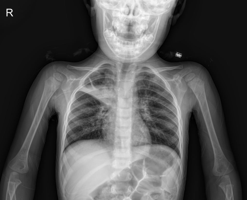
- X-ray Interpretation: The PA chest X-ray revealed a linear fissure area in the right upper zone. This finding raised suspicion for pneumonia, particularly given the diminished breath sounds in that area during physical examination.
Incidental Metabolic Findings
During the initial assessment, the patient’s blood glucose levels were significantly elevated:
- Capillary Blood Glucose (CBG): 350 mg/dL
- Point-of-Care Blood Glucose (POCT): 444 mg/dL
- Lactate: 6.20 mmol/L
Due to these findings, the patient was admitted for intravenous hydration and close monitoring. Urinalysis showed ketones 2+, further supporting the suspicion of newly diagnosed diabetes mellitus. Further investigations for new-onset diabetes were initiated, including insulin antibodies, GAD antibodies, thyroid function tests (anti-TPO, anti-thyroglobulin, T4, T3, TSH), tissue transglutaminase IgA, IgA turbidimetric assay, C-peptide, and insulin levels. During observation, the patient’s POCT blood glucose levels normalized and remained stable. The patient was subsequently transferred to the pediatric ward for further evaluation and management.
Patient History & Family Background
- Parents: Not related, healthy. Mother reported kidney enlargement during both pregnancies with a history of unknown antibiotic use.
- Sibling: 7-year-old older brother, healthy.
- Birth History: 41 weeks gestational age, normal spontaneous delivery. No history of neonatal intensive care unit (NICU) admission. Routine vaccinations were not completed.
- Home Environment: No humidity, no pets.
Hospital Course (Day 1)
The patient was admitted with preliminary diagnoses of possible lung fissure (pneumonia) and for monitoring of hyperglycemia.
- Current Status: Weight: 12 kg (0.54 m² BSA), Height: 99 cm.
- Physical Examination:
- General condition: Moderate.
- Vitals: Afebrile. SpO2: 96-99% on room air. Heart rate: 110-130 bpm.
- Skin: Normal, no rash.
- Head-Neck: Normal, no cervical lymphadenopathy.
- Oropharynx: Normal.
- Respiratory System: Harsh breath sounds bilaterally, rales (+) in the right lung, no rhonchi, no wheezing, no intercostal retractions. Mild abdominal breathing and tachypnea.
- Cardiovascular System: S1+ S2+, no extra sounds, no murmurs.
- Gastrointestinal System: Abdomen soft, no defense, no rebound tenderness, bowel sounds present.
- Genitourinary System: Externally male, no major urogenital anomalies, urine output present.
- Neuromuscular System: GCS: 15, conscious, pupils isocoric, light reflex +/+, no meningeal irritation signs.
- Extremities: No deformities.
Treatment Plan
- Ampicillin-sulbactam: With proper doses (Day 1).
- Hypertonic saline: With proper doses via nebulization.
- Budesonide: With proper doses via nebulization.
- Epinephrine: With proper doses via nebulization (Day 1).
- 5% Dextrose + 0.9% Normal Saline (with 10 mL KCl added to every 500 mL if diuresis present): Infusion rate with proper doses (due to poor appetite).
- Point-of-care blood glucose monitoring: Every 4 hours.
Please note: Drug types and dosages should be verified according to your country’s relevant pharmaceutical dosage guidelines, or by consulting a licensed physician in the relevant department.
Further Investigations & Management Plan
- Urine culture monitoring.
- Anemia panel to be drawn during blood collection or IV line insertion (due to weight below CDC percentile and low MCV).
- Follow-up on results for anti-insulin antibody, anti-GAD antibody, anti-TPO, anti-thyroglobulin antibody, T4, T3, TSH, tissue transglutaminase IgA, IgA turbidimetric, C-peptide, and insulin levels.
Conclusion
This case highlights the importance of thorough clinical and radiological assessment in pediatric patients presenting with respiratory symptoms. The presence of a linear fissure on chest X-ray in a child with a barking cough and diminished breath sounds warrants consideration of pneumonia, even in the context of typical croup symptoms. Furthermore, the incidental finding of significant hyperglycemia underscores the need for comprehensive initial workups and vigilance for unexpected systemic conditions, such as new-onset diabetes.
This content is for educational and reference purposes only and should not be used as a basis for diagnosis or treatment. Always consult with a qualified healthcare professional for any medical concerns.

