The microscopic examination of blood vessels offers a window into the intricate cellular and tissue architecture that sustains the circulatory system. This image, captured under a microscope, highlights the tunica intima, tunica media, tunica adventitia, and endothelial cells, revealing the structural adaptations that enable arteries, veins, and capillaries to perform their unique roles.
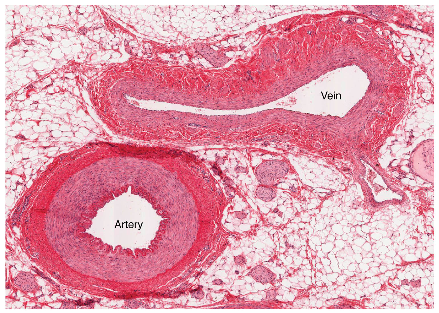
Tunica intima Tunica intima is the innermost layer visible under the microscope, featuring a single layer of endothelial cells that line the vessel lumen. This layer reduces friction and prevents clot formation, with a subendothelial layer and elastic lamina adding support in larger vessels.
Tunica media Tunica media appears as the middle layer, composed of smooth muscle cells and elastic fibers, varying in thickness depending on the vessel type. In arteries, this layer is prominent with elastic lamellae, while in veins, it is thinner, reflecting their pressure differences.
Tunica adventitia Tunica adventitia forms the outermost layer, consisting of connective tissue and collagen fibers that anchor the vessel to surrounding tissues. This layer contains vasa vasorum, which supply nutrients to the vessel wall, and is more pronounced in veins.
Endothelial cells Endothelial cells line the tunica intima, appearing as a continuous sheet of flattened cells under the microscope. These cells regulate blood flow, prevent clotting by releasing nitric oxide, and facilitate nutrient exchange, playing a critical role in vascular health.
The Role of Microscopic Layers in Blood Vessels
This exploration uncovers how each layer contributes to vascular function at a cellular level. The microscopic view provides clarity on how these structures support circulation.
- Tunica Intima Function: The endothelial cells form a barrier that controls substance exchange between blood and tissues. The elastic lamina in arteries enhances flexibility under pressure.
- Tunica Media Role: Smooth muscle cells contract or relax to adjust vessel diameter, influenced by hormonal signals. Elastic fibers in arteries store energy, aiding pulse smoothing.
- Tunica Adventitia Support: The collagen network provides tensile strength, preventing vessel rupture. Vasa vasorum ensure oxygen delivery to the outer layers of larger vessels.
- Endothelial Cell Activity: These cells secrete factors like prostacyclin to inhibit platelet aggregation. They also respond to shear stress, adjusting vessel tone.
Anatomical Details Under the Microscope
The microscopic image reveals the fine structure of blood vessel layers with precision. The magnification highlights cellular organization and tissue density.
This detailed view allows for a deeper understanding of vascular anatomy. It serves as a foundation for studying tissue health and pathology.
- Tunica Intima Composition: The endothelial layer is supported by a basement membrane, with the subendothelial layer varying in thickness. Elastic lamellae are more visible in arterial sections.
- Tunica Media Thickness: The layer shows concentric smooth muscle bands in arteries, with elastic fibers interspersed. In veins, the muscle layer is sparse, with more connective tissue.
- Tunica Adventitia Variation: Collagen fibers dominate this layer, appearing as dense networks under the microscope. Vasa vasorum are small, branching structures within this region.
- Endothelial Cell Morphology: These cells appear elongated in arteries due to high pressure, while more rounded in veins. Their nuclei are visible, indicating active cellular function.
Physiological Functions of Microscopic Structures
The physiological roles of these layers are evident at the microscopic level. Their design supports the diverse needs of the circulatory system.
Each component plays a specific role in maintaining blood flow. This functionality underpins the body’s overall vascular health.
- Blood Pressure Regulation: The tunica media’s elastic fibers in arteries absorb systolic pressure, smoothing flow. This mechanism protects downstream capillaries.
- Nutrient Exchange: Endothelial cells in the tunica intima facilitate oxygen and glucose diffusion in capillaries. This exchange is critical for tissue metabolism.
- Venous Return: The thin tunica media in veins relies on external compression, with valves aiding flow. The adventitia supports this passive movement.
- Clot Prevention: Endothelial cells release anticoagulants like heparin, preventing thrombosis. This action maintains vessel patency.
Comparative Anatomy Across Vessel Types
The microscopic view allows comparison between arterial, venous, and capillary structures. These differences are tailored to their specific circulatory roles.
The image underscores the variation in layer prominence. This contrast is key to understanding vascular diversity.
- Arterial vs. Venous Layers: Arteries show a thick tunica media with elastic lamellae, while veins have a thicker tunica adventitia. This reflects their pressure and flow differences.
- Capillary Simplicity: Capillaries reduce to a single endothelial layer, lacking media and adventitia. This thinness maximizes exchange efficiency.
- Elasticity Contrast: The tunica media in arteries contains more elastic fibers, visible as dark bands. Veins rely on collagen, appearing lighter under the microscope.
- Staining Techniques: Hematoxylin stains nuclei blue, while eosin highlights elastic and collagen fibers. This differentiation aids in identifying vessel types.
Clinical and Research Perspectives
Microscopic views of blood vessels offer valuable insights for medical practice. The cellular structure is a focus for studying vascular diseases and health.
Advances in microscopy and histology enhance these studies, providing diagnostic tools. These efforts support innovative treatment approaches.
- Atherosclerosis Detection: Plaque in the tunica intima appears as lipid deposits under the microscope. Early identification guides lipid-lowering therapy.
- Varicose Vein Assessment: Thinning of the tunica media and valve damage are visible in affected veins. Microscopic analysis informs compression treatment.
- Endothelial Dysfunction: Damaged endothelial cells show irregular shapes, indicating inflammation. This finding directs antihypertensive strategies.
- Therapeutic Innovations: Targeting endothelial nitric oxide production treats hypertension. Stem cell research explores regenerating vessel layers.
In conclusion, this image of blood vessels under the microscope provides a detailed look at the tunica intima, tunica media, tunica adventitia, and endothelial cells, revealing their critical roles in circulation. These microscopic insights not only enhance our understanding of vascular anatomy but also support advancements in diagnosing and treating circulatory conditions.

