The brain relies on a sophisticated network of arteries to receive a continuous supply of oxygenated blood, essential for its complex functions. This inferior view diagram showcases the arterial circle, known as the circle of Willis, which interconnects major arteries to ensure consistent cerebral perfusion and resilience against vascular interruptions.
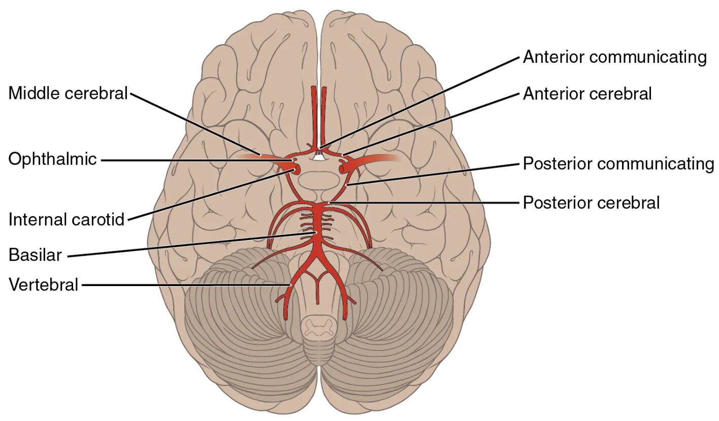
Internal carotid arteries These arteries deliver oxygenated blood from the common carotid arteries to the brain. They branch into the anterior and middle cerebral arteries within the cranial cavity.
Anterior cerebral arteries Arising from the internal carotid arteries, they supply blood to the frontal lobes and medial aspects of the brain. They connect via the anterior communicating artery to form part of the circle of Willis.
Anterior communicating artery This short vessel links the two anterior cerebral arteries. It allows blood to flow between the left and right sides, enhancing collateral circulation.
Middle cerebral arteries Branching from the internal carotid arteries, they provide blood to the lateral aspects of the brain, including much of the cerebral cortex. They are critical for functions like language and motor control.
Posterior cerebral arteries These arteries arise from the basilar artery and supply the occipital lobes and parts of the temporal lobes. They connect to the internal carotid system via the posterior communicating arteries.
Posterior communicating arteries Linking the posterior cerebral arteries to the internal carotid arteries, these vessels complete the circle of Willis. They ensure backup blood flow if one pathway is compromised.
Basilar artery Formed by the fusion of the two vertebral arteries, it runs along the brainstem. It gives rise to the posterior cerebral arteries, supplying the lower brain regions.
Vertebral arteries Originating from the subclavian arteries, they ascend through the cervical vertebrae to join as the basilar artery. They provide blood to the brainstem and cerebellum.
Circle of Willis This ring-like structure at the base of the brain connects the internal carotid and vertebral systems. It serves as a safety net, redistributing blood if an artery is blocked.
Anatomy of the Circle of Willis
The circle of Willis forms a critical junction for cerebral blood supply. Its circular arrangement enhances the brain’s ability to adapt to changes.
- The internal carotid arteries enter the circle, branching into anterior and middle cerebral arteries.
- The anterior communicating artery bridges the two sides for symmetry.
- Posterior communicating arteries integrate the vertebral system into the network.
- The basilar artery, fed by vertebral arteries, supports the posterior circulation.
- This structure’s redundancy protects against ischemic damage.
Role of Major Cerebral Arteries
The anterior cerebral arteries and their counterparts are vital for specific brain regions. Their distribution ensures comprehensive coverage.
- Anterior cerebral arteries nourish the frontal lobes, influencing cognition.
- Middle cerebral arteries supply the lateral cortex, critical for speech.
- Posterior cerebral arteries feed the visual cortex in the occipital lobes.
- These arteries adjust flow based on metabolic demands.
- Variations in their paths can affect surgical planning.
Contribution of Vertebral and Basilar Systems
The vertebral arteries and basilar artery support the posterior brain regions. Their upward path ensures robust supply to the brainstem.
- Vertebral arteries travel through the cervical spine, merging at the skull base.
- The basilar artery runs along the brainstem, branching to the posterior cerebrals.
- This system supplies the cerebellum, vital for coordination.
- Collateral flow here prevents deficits from vertebral occlusion.
- The pathway’s vulnerability requires careful monitoring.
Physiological Importance of the Arterial Circle
The circle of Willis maintains blood pressure and flow to the brain under varying conditions. Its design offers a protective mechanism.
- It redistributes blood if one artery narrows or blocks.
- The circle equalizes pressure between carotid and vertebral systems.
- Oxygen delivery remains stable during postural changes.
- Metabolic activity in the brain drives local flow adjustments.
- This adaptability is key to preventing strokes or hypoxia.
Clinical Relevance of Brain Arteries
Understanding the anatomy aids in diagnosing and treating cerebral vascular issues. The circle’s structure guides medical interventions.
- Aneurysms in the anterior communicating artery are common and risky.
- Blockage of middle cerebral arteries can cause hemiplegia.
- Posterior cerebral artery issues may lead to visual field loss.
- Vertebral artery dissection is a concern in neck trauma.
- Angiography visualizes the circle to plan treatments like stenting.
The circle of Willis, with its interconnected internal carotid arteries, vertebral arteries, and other branches, ensures the brain receives a steady supply of oxygenated blood. This resilient network supports cognitive and motor functions, offering valuable insights into cerebral health and potential therapeutic strategies.

