The arterial system is a vital component of the circulatory network, designed to transport oxygenated blood under high pressure from the heart to the body’s tissues. This image offers a detailed sectional view of an artery, highlighting the tunica intima, tunica media, and tunica adventitia, which together provide the strength and elasticity needed to withstand pulsatile blood flow.
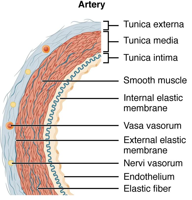
Tunica intima Tunica intima is the innermost layer of the artery, featuring a single layer of endothelial cells that reduce friction and prevent clot formation. This layer includes a subendothelial layer and an internal elastic lamina, which enhances the artery’s ability to stretch and recoil with each heartbeat.
Tunica media Tunica media forms the thick middle layer, composed of smooth muscle cells and multiple elastic lamellae that regulate vessel diameter and absorb pressure. This robust layer enables the artery to handle the high systolic pressure, up to 120 mmHg, while maintaining structural integrity.
Tunica adventitia Tunica adventitia is the outer layer, consisting of connective tissue and collagen fibers that anchor the artery to surrounding tissues. This layer contains the vasa vasorum, small vessels that supply nutrients to the arterial wall, especially important in larger arteries.
The Role of Arterial Layers in Circulation
This analysis reveals how each layer contributes to the artery’s function. The specialized design ensures efficient blood delivery under high pressure.
- Tunica Intima Function: The endothelium releases nitric oxide, promoting vasodilation and preventing platelet aggregation. The internal elastic lamina provides flexibility, accommodating the pulse wave.
- Tunica Media Role: The smooth muscle allows vasoconstriction to regulate blood flow during stress or exercise. Elastic fibers store energy during systole, releasing it during diastole to maintain pressure.
- Tunica Adventitia Support: This layer stabilizes the artery, preventing rupture under pressure. The vasa vasorum ensures oxygen and nutrients reach the outer wall layers.
Anatomical Details of Arterial Structure
The sectional view showcases the artery’s layered construction, offering insight into its resilience. The thickness of each layer reflects its adaptation to high-pressure environments.
The image’s cross-sectional perspective highlights the artery’s robust design. This understanding is essential for studying vascular health and function.
- Tunica Intima Composition: The endothelial layer is supported by a thin subendothelial connective tissue and the internal elastic lamina. This structure varies in thickness depending on the artery’s size and location.
- Tunica Media Thickness: This layer contains up to 40 layers of elastic lamellae in large arteries like the aorta, interspersed with smooth muscle. The density of elastic tissue decreases in smaller arteries.
- Tunica Adventitia Variation: The outer layer is thinner than the media but rich in collagen, providing tensile strength. The vasa vasorum becomes more prominent in larger arteries to support deeper tissue layers.
- Elastic Lamellae: These concentric sheets within the tunica media enhance elasticity, crucial for large arteries. They allow the vessel to expand and contract with each cardiac cycle.
Physiological Functions of Arterial Layers
The physiological roles of these layers are tailored to handle high-pressure blood flow. Their design ensures effective oxygen and nutrient delivery to tissues.
Each layer plays a specific role in maintaining circulation. This functionality supports the body’s metabolic demands under varying conditions.
- Blood Pressure Management: The tunica media’s elastic fibers absorb the systolic peak, smoothing the pressure wave. This prevents damage to smaller downstream vessels.
- Vasomotor Control: Smooth muscle in the tunica media responds to neurotransmitters like acetylcholine, adjusting vessel diameter. This regulation balances blood distribution during activity.
- Oxygen Delivery: Arteries carry oxygenated blood rich in nutrients like glucose and amino acids. The tunica intima’s smoothness ensures efficient transport to capillary beds.
- Pulse Propagation: The elastic recoil of the tunica media and intima propagates the pulse wave, aiding peripheral perfusion. This mechanism is vital for maintaining systemic pressure.
Comparative Anatomy with Other Vessels
The arterial structure differs significantly from veins and capillaries. This sectional view emphasizes the artery’s unique adaptations to its role.
The image underscores the artery’s distinct layering compared to veins. This contrast is key to understanding circulatory dynamics.
- Arterial vs. Venous Walls: Arteries have a thicker tunica media due to high pressure, while veins rely on a thicker adventitia and valves. This difference reflects their respective functions.
- Transition to Capillaries: The tunica media thins as arteries branch into arterioles, reducing to a single endothelial layer in capillaries. This transition maximizes exchange surfaces.
- Elasticity Gradient: Large elastic arteries like the aorta have more elastic lamellae than muscular arteries. This gradient supports varying pressure needs across the arterial tree.
- Histological Features: Staining reveals dense elastic fibers in the tunica media, contrasting with the collagen dominance in veins. This aids in microscopic identification.
Clinical and Research Perspectives
Insights from arterial sectional views inform medical practice and research. The layered structure is a focus for studying cardiovascular health and disease.
Advances in imaging and histology enhance these studies, offering diagnostic tools. These efforts support innovative treatment strategies.
- Atherosclerosis Development: Plaque buildup in the tunica intima narrows the lumen, increasing risk. Sectional analysis guides early intervention like stenting.
- Hypertension Impact: Thickened tunica media in response to chronic high pressure can stiffen arteries. Microscopic views assess this remodeling.
- Surgical Considerations: Arterial grafting requires matching layer thickness, especially the tunica media. This ensures graft durability.
- Therapeutic Advances: Targeting tunica media smooth muscle with drugs like beta-blockers treats hypertension. Stem cell research explores regenerating arterial layers.
In conclusion, this image of an arterial sectional view provides a detailed look at the tunica intima, tunica media, and tunica adventitia, highlighting their critical roles in high-pressure blood transport. These anatomical insights not only enhance our understanding of arterial function but also support advancements in diagnosing and treating cardiovascular conditions.

