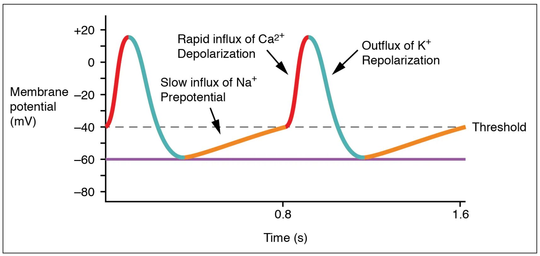
The sinoatrial (SA) node, as the heart’s natural pacemaker, generates electrical impulses that initiate each heartbeat, a process vividly illustrated in this diagram. This image details the prepotential, threshold, rapid depolarization, and repolarization phases, highlighting the unique absence of a resting potential and the role of sodium ion influx in driving spontaneous activity. Exploring this diagram provides a clear understanding of how the SA node sustains the heart’s rhythmic contractions.
Labelled Parts Explanation
- Prepotential The prepotential is a gradual depolarization phase caused by a slow influx of sodium ions, bringing the membrane toward the threshold. It accounts for the SA node’s spontaneous activity, initiating the action potential without a stable resting potential.
- Threshold The threshold is the critical membrane potential at which rapid depolarization begins, triggered by the prepotential’s sodium influx. It marks the point where the SA node activates, leading to atrial contraction.
- Rapid depolarization The rapid depolarization phase involves a swift influx of sodium ions, sharply increasing the membrane potential after the threshold is reached. This phase generates the electrical impulse that spreads across the atria to initiate systole.
- Repolarization The repolarization phase restores the membrane potential to its prepotential state through potassium ion efflux, following rapid depolarization. It prepares the SA node for the next cycle of spontaneous activity.
Anatomical Overview of the SA Node Action Potential
The SA node’s action potential is a unique electrical event that drives the cardiac cycle. This diagram illustrates the phases that ensure continuous heart rhythm.
- The prepotential gradually depolarizes the membrane, setting the stage for impulse generation.
- The threshold serves as the trigger point, initiating the rapid depolarization phase.
- The repolarization phase resets the SA node, enabling its pacemaker function.
- The absence of a resting potential distinguishes the SA node from other cardiac cells.
This process underpins the heart’s ability to beat autonomously.
Mechanism of Prepotential and Threshold
The prepotential and threshold are the foundation of the SA node’s pacemaker activity. Their dynamics initiate each heartbeat.
- The prepotential results from a slow, funny current of sodium ions entering the cell.
- This gradual rise brings the membrane to the threshold, where voltage-gated sodium channels open.
- The process occurs without a stable resting potential, reflecting the SA node’s unique physiology.
- Autonomic modulation can adjust the prepotential slope, altering heart rate.
This mechanism ensures spontaneous and adjustable rhythm.
Process of Rapid Depolarization and Repolarization
Rapid depolarization and repolarization complete the action potential cycle. These phases coordinate atrial contraction.
- The rapid depolarization phase sees a surge of sodium ions, creating a steep voltage spike.
- The repolarization phase follows as potassium ions exit, returning the membrane to its prepotential state.
- This sequence generates the electrical signal that spreads via internodal pathways.
- Calcium ions also contribute to the plateau phase, though less prominent in the SA node.
These stages are critical for effective atrial systole.
Physiological Significance of the SA Node Action Potential
The SA node’s action potential supports the heart’s continuous operation. Its design optimizes rhythm regulation.
- The prepotential’s spontaneous nature allows the SA node to act as the primary pacemaker.
- The threshold and rapid depolarization ensure timely atrial activation.
- The repolarization phase prepares the node for the next cycle, maintaining rhythm.
- This process adapts to physiological demands through neural and hormonal influences.
The SA node’s activity is essential for cardiovascular stability.
Ionic Basis of the Action Potential
The action potential relies on specific ion movements across the SA node membrane. These shifts drive electrical changes.
- The prepotential is driven by a slow influx of sodium ions via funny channels.
- The rapid depolarization involves a rapid sodium influx, amplified by calcium entry.
- The repolarization phase depends on potassium efflux to reset the membrane.
- The lack of a resting potential reflects the SA node’s pacemaker current.
This ionic dance sustains the heart’s rhythmic beating.
Clinical Relevance of the SA Node Action Potential
Understanding the SA node’s action potential aids in diagnosing rhythm disorders. These phases are key clinical markers.
- Abnormal prepotential slopes can lead to sick sinus syndrome, causing bradycardia.
- Failure to reach the threshold may result in pauses or asystole.
- Altered rapid depolarization or repolarization can contribute to tachycardia.
- Electrocardiograms assess these phases to guide treatment like pacing.
This knowledge supports effective management of cardiac arrhythmias.
Conclusion
The action potential at the SA node diagram offers a detailed look at the electrical process that initiates each heartbeat. By examining the prepotential, threshold, rapid depolarization, and repolarization phases, one gains insight into how the SA node maintains the heart’s rhythm without a resting potential. This understanding serves as a foundation for studying cardiovascular physiology and addressing related health issues, encouraging further exploration of the heart’s intricate electrical design and its critical role in sustaining life.

