Carotid ultrasonography has emerged as a cornerstone in non-invasive vascular imaging, providing a direct window into the systemic health of the arterial tree. The Common Carotid Artery (CCA), particularly its proximal segment, is a frequent site for assessing the early stages of structural vascular changes. By measuring the Intima-Media Thickness (IMT), clinicians can detect the silent progression of subclinical atherosclerosis before any overt symptoms occur. This measurement acts as a surrogate marker for generalized vascular aging and is strongly correlated with the risk of future myocardial infarction and stroke. Through high-frequency ultrasound, the intricate layers of the vessel wall—the intima, media, and adventitia—can be visualized with sub-millimetric precision, allowing for a proactive approach to cardiovascular health management. Understanding the anatomical relationships and the technical nuances of these measurements is essential for the modern clinician in the field of preventive cardiology.
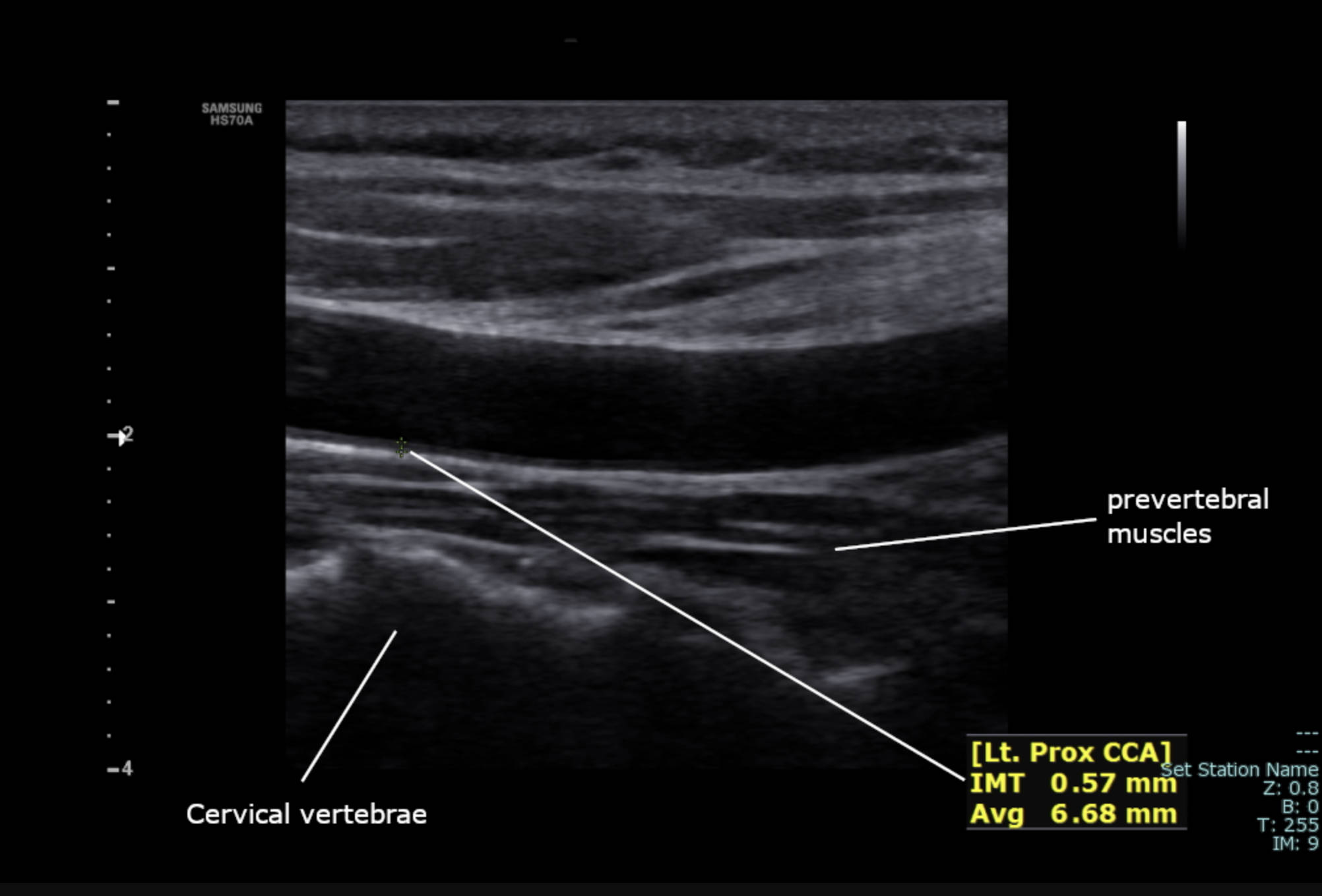
prevertebral muscles: These are the deep muscular structures of the neck located just anterior to the spine, appearing on ultrasound as a striated hypoechoic region. They serve as a vital posterior anatomical landmark for identifying the carotid sheath and ensuring the ultrasound probe is properly oriented.
Cervical vertebrae: These bony segments of the neck appear as a bright, echogenic line with dark posterior acoustic shadowing beneath the muscles. Their presence in the image confirms the depth of the scan and helps the sonographer navigate the complex cross-sectional anatomy of the cervical region.
[Lt. Prox CCA]: This designation identifies the scan as the Left Proximal Common Carotid Artery, specifying both the side of the body and the longitudinal segment of the vessel. Precise labeling is mandatory in clinical practice to track changes in the arterial wall over multiple follow-up examinations.
IMT 0.57 mm: This is the measured Intima-Media Thickness, representing the combined thickness of the innermost two layers of the arterial wall. A value of 0.57 mm is generally considered within the healthy range for an adult, reflecting a low immediate burden of structural arterial aging.
Avg 6.68 mm: This numerical value represents the average luminal diameter of the carotid artery at the measurement site. Monitoring the diameter in conjunction with wall thickness provides a comprehensive view of the hemodynamic capacity and structural integrity of the vessel.
Understanding Carotid Intima-Media Thickness (IMT)
Carotid Intima-Media Thickness, commonly referred to as IMT, is a validated measurement of the double-layer thickness of the innermost two segments of the arterial wall: the intima and the media. On a high-resolution intima-media thickness scan, these layers appear as two parallel echogenic lines separated by a hypoechoic space. The measurement is typically taken at the far wall of the common carotid artery, as this has been shown to be the most reproducible and representative site for systemic vascular health. As individuals age or are exposed to metabolic stressors, these layers naturally thicken, but excessive thickening serves as an early warning sign of systemic arterial disease.
The clinical utility of IMT lies in its ability to detect pathology long before a discrete plaque is formed or a lumen becomes narrowed. It is considered a continuous variable; even within what is statistically defined as a “normal” range, higher values are often associated with a higher cardiovascular risk profile. For researchers and clinicians alike, IMT provides a quantitative way to assess the efficacy of interventions, such as lipid-lowering therapies or lifestyle changes, by observing the stabilization or regression of the arterial wall thickness over months or years.
The Role of Doppler Ultrasound in Vascular Assessment
Modern Doppler ultrasound systems, such as the Samsung HS70A utilized in this image, combine B-mode (brightness mode) imaging with Doppler physics to provide both structural and functional data. While the B-mode image allows for the precise measurement of the IMT as seen here, Doppler technology allows clinicians to measure the velocity and direction of blood flow within the vessel. This is particularly important in cases where IMT is high, as it helps determine if the thickening is causing significant hemodynamic turbulence or obstruction. The proximal common carotid artery is an ideal site for this because of its relatively straight course and accessibility.
Technological advancements have made these measurements incredibly precise. Semi-automated software can now track the intima-media interface along a segment of several centimeters, calculating the average thickness to reduce human error. This ensures that the “0.57 mm” seen in the measurement is not just a single point of data, but a statistically significant representation of the vessel’s health. The non-invasive nature of ultrasound, combined with its lack of ionizing radiation, makes it the preferred tool for screening asymptomatic populations who may be at risk for future cardiac events.
Interpreting Clinical Measurements and Ranges
In clinical practice, interpreting an IMT value requires context, primarily age and gender. While a measurement of 0.57 mm is excellent for a 60-year-old, it might be at the upper limit for a very young adult. Generally, values below 0.6 mm are considered very low risk, while those exceeding 0.9 mm or 1.0 mm are often indicative of significantly increased risk and the likely presence of atherosclerosis elsewhere in the body. If the thickness exceeds 1.5 mm, it is usually reclassified from “thickening” to a “focal plaque,” which carries a different set of clinical management guidelines.
The average diameter of the vessel, in this case 6.68 mm, is also a useful metric. While diameter varies based on body size and blood pressure, significant deviations can indicate arterial remodeling. In hypertensive patients, the artery might dilate (eccentric remodeling) or the wall might thicken (concentric remodeling) to compensate for the increased wall tension. By viewing both the IMT and the average diameter together, a physician can gain a deeper understanding of how the patient’s vascular system is adapting to their specific physiological environment.
Anatomical Landmarks and Scanning Technique
Successful carotid imaging depends heavily on the identification of neighboring structures. As seen in the micrograph, the prevertebral muscles and the cervical vertebrae provide a steady backdrop. Identifying these structures ensures that the sonographer is scanning at the correct depth and that the carotid artery is properly distinguished from the more superficial internal jugular vein. The carotid artery is typically characterized by its thicker walls, pulsatality, and lack of respiratory variation, whereas the vein is highly compressible and changes size with the patient’s breathing.
- Longitudinal View: The probe is oriented along the length of the neck to see the vessel as a tube, as shown in the image. This is necessary for measuring IMT.
- Transverse View: The probe is turned 90 degrees to see the vessel in cross-section, which is better for identifying the circumferential distribution of plaque.
- Angle of Insonation: The ultrasound beam must be perpendicular to the vessel wall to get the clearest definition of the intima-media layers.
- Gain Settings: Proper adjustment of the machine’s gain is essential to differentiate the dark lumen from the gray vessel wall.
Significance in Preventive Medicine
The primary goal of measuring carotid IMT is prevention. By identifying individuals with accelerated vascular aging, doctors can implement primary prevention strategies more aggressively. This might involve stricter blood pressure targets, earlier initiation of statin therapy, or more intensive smoking cessation counseling. Because atherosclerosis is a systemic process, the state of the carotid artery is often a “proxy” for the state of the coronary arteries. If the carotids are healthy, the risk of a silent coronary blockage is statistically lower.
Furthermore, this imaging modality is invaluable in educating patients. Seeing a visual representation of their own arterial wall can be a powerful motivator for lifestyle changes. A measurement like 0.57 mm can provide reassurance, while a thicker measurement can serve as a wake-up call. In an era where personalized medicine is becoming the standard, carotid IMT provides a patient-specific biomarker that moves beyond general risk calculators based on population averages.
Conclusion
The ultrasound assessment of the left proximal common carotid artery represents a sophisticated intersection of physics, anatomy, and clinical medicine. Through the precise measurement of the intima-media thickness and the luminal diameter, clinicians are gifted with a powerful tool for predicting and preventing cardiovascular catastrophes. The image provided illustrates a healthy vascular profile, but the underlying technology is what allows us to catch disease in its infancy. As ultrasound hardware and software continue to evolve, our ability to map the landscape of the human arterial system will only improve, leading to better outcomes and longer, healthier lives for patients worldwide.

