Observing Euglena under the microscope reveals the dynamic life of a versatile mixotrophic protist that combines photosynthetic and heterotrophic capabilities within a single cell. This elongated, green organism, commonly found in freshwater ponds and ditches, displays striking internal structures and active behaviors that make it a favorite subject in biology laboratories worldwide. The high-resolution microscopic image captures key diagnostic features that allow students, educators, and researchers to appreciate the sophisticated adaptations enabling Euglena to thrive in environments with fluctuating light and nutrient availability.
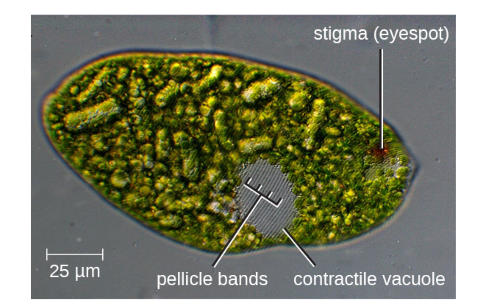
Stigma (eyespot) is the prominent orange-red spot visible near one end of the cell. It functions as a shading device for the photoreceptor, allowing Euglena to detect the direction of light and perform phototaxis to position itself optimally for photosynthesis.
Pellicle bands are the visible helical or longitudinal stripes running along the cell surface. These proteinaceous strips form the flexible pellicle that provides structural support while permitting the characteristic euglenoid movement (metaboly) used for shape changes and locomotion.
Contractile vacuole is the clear, spherical structure often visible near the anterior or central region. It collects excess water from the cytoplasm and periodically contracts to expel fluid through a pore, maintaining osmotic balance in the hypotonic freshwater environment.
25 μm scale bar indicates the microscopic scale of the specimen. It confirms that Euglena is a relatively large protist, easily observable under standard light microscopy and ideal for detailed live-cell observation in educational settings.
Live Microscopic Appearance of Euglena
Under the light microscope, living Euglena appears as a bright green, elongated cell with a distinct orange-red stigma near the anterior end. The pellicle bands give the surface a striated texture, and the cell often exhibits graceful euglenoid movement, changing shape from elongated to more rounded forms. The contractile vacuole can be seen pulsing at regular intervals, while the green chloroplasts fill much of the cytoplasm, sometimes appearing as distinct disc-like structures. When actively swimming, the anterior flagellum propels the cell forward in a characteristic spiral path.
Key Visible Structures and Their Functions
The microscopic image clearly displays several diagnostic features. The stigma stands out as a bright orange-red dot, essential for light detection. The pellicle bands create a patterned surface texture that reflects the underlying protein strips. The contractile vacuole appears as a clear circular area, often located near the reservoir at the anterior end. These visible elements allow quick identification of Euglena among other freshwater protists and provide immediate insight into its physiological activities during live observation.
Phototaxis and Light Sensing
The stigma and associated photoreceptor enable Euglena to perform sophisticated phototaxis. As the cell rotates while swimming, the stigma periodically shades the photoreceptor, generating a signal that the organism uses to steer toward light sources. This behavior ensures optimal positioning for photosynthesis and demonstrates how a single cell can integrate sensory information to produce directed movement without a nervous system.
Pellicle and Euglenoid Movement
The pellicle bands visible in the image represent the flexible outer covering that allows Euglena to perform metaboly. This crawling or squirming movement enables the organism to navigate through viscous environments or narrow spaces when flagellar swimming is insufficient. The ability to change shape rapidly is a key adaptation for survival in variable aquatic habitats and distinguishes Euglena from many other flagellated protists.
- Pellicle provides both support and flexibility for shape changes.
- Euglenoid movement supplements flagellar propulsion.
- Contractile vacuole activity is easily observed in live specimens.
These features make Euglena particularly engaging for live microscopy sessions.
Metabolic Versatility Demonstrated in Live Cells
Live Euglena cells display clear evidence of their mixotrophic nature. In bright light, the intense green color from chloroplasts indicates active photosynthesis. When kept in darkness for extended periods, cells gradually lose their green pigmentation as chloroplasts reduce activity, switching to heterotrophic nutrition. The presence of the contractile vacuole in the image highlights ongoing osmoregulation, a constant physiological demand in freshwater environments.
Educational Value of Microscopic Observation
Euglena is one of the most popular organisms for introductory microscopy labs because students can observe living cells performing multiple vital functions simultaneously. The visible stigma, pellicle bands, and contractile vacuole provide immediate points of interest, while the active swimming and shape changes maintain student engagement. Comparing Euglena with other protists such as Paramecium or Amoeba helps illustrate the diversity of eukaryotic adaptations and nutritional strategies.
Research Applications and Broader Significance
Beyond education, Euglena serves as a valuable model organism in research on photosynthesis, circadian rhythms, flagellar biology, and metabolic regulation. Its large size and ease of cultivation facilitate advanced imaging techniques and genetic studies. Research on Euglena has contributed to understanding chloroplast evolution and has potential applications in biotechnology, including the production of nutritional supplements and biofuels from its paramylon reserves.
Ecological Context and Identification Tips
In natural freshwater ecosystems, Euglena often appears in nutrient-rich or slightly polluted waters, sometimes forming green blooms. Under the microscope, the combination of the red stigma, green chloroplasts, and pellicle bands allows reliable identification even among other green flagellates. The 25 μm scale bar in the image helps students appreciate its relatively large size compared to many other protists.
Conclusion: The Dynamic World of Living Euglena
The microscopic view of Euglena beautifully captures the elegance and functional sophistication of a single-celled organism. From the distinctive stigma and pellicle bands to the active contractile vacuole, every visible feature reflects adaptations for survival in dynamic aquatic environments. As both an educational tool and a research model, living Euglena continues to inspire fascination with the complexity achievable in unicellular life, bridging basic observation with deeper insights into eukaryotic cell biology, metabolism, and behavior.

