Mycobacterium tuberculosis, the causative agent of tuberculosis, remains one of the world’s most persistent infectious threats, affecting millions annually and requiring specialized laboratory techniques for reliable detection. Culture on solid media continues to serve as a cornerstone for definitive diagnosis, drug susceptibility testing, and epidemiological tracking despite the rise of molecular methods. The provided image showcases the distinctive growth pattern of M. tuberculosis on Löwenstein-Jensen (LJ) agar, a traditional egg-based medium optimized for this slow-growing acid-fast bacillus, offering visual insight into its characteristic colony features after weeks of incubation.
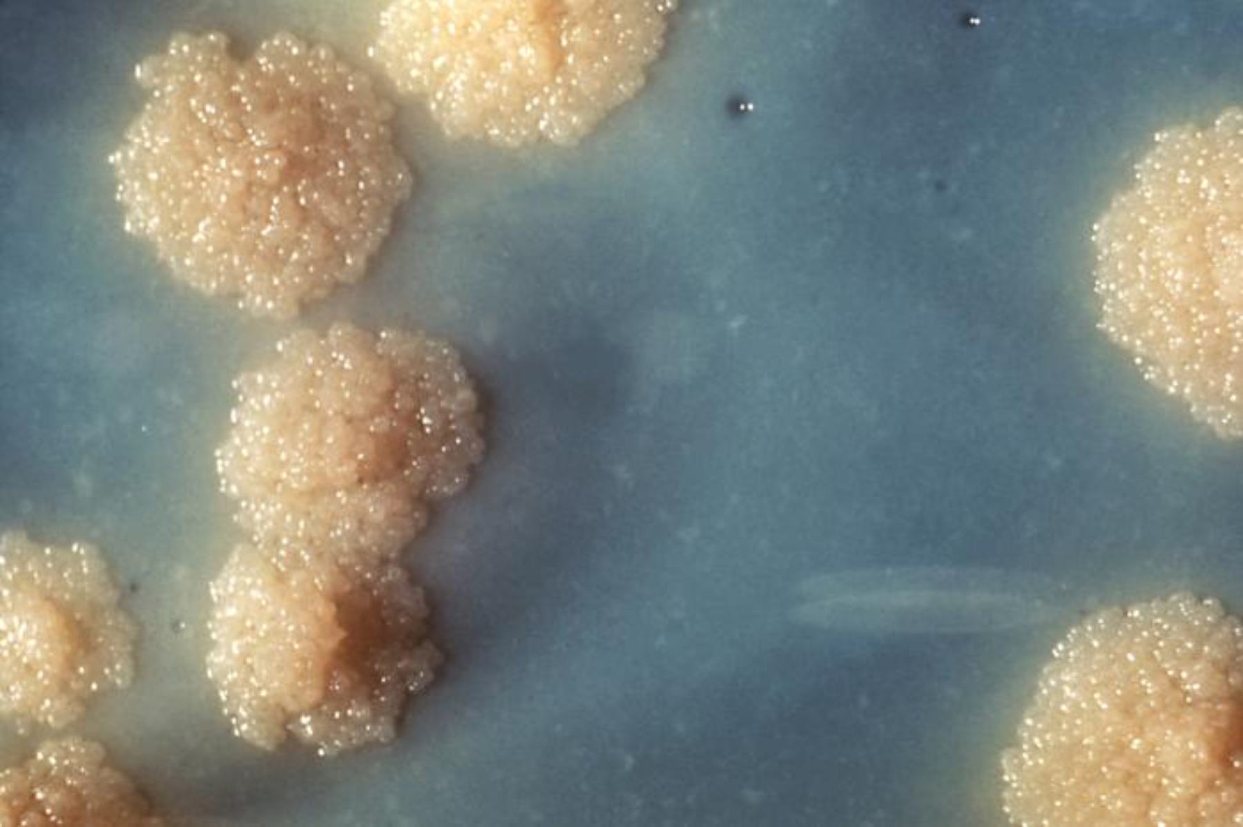
Löwenstein-Jensen agar is the selective solid medium depicted in the image with its characteristic bluish-green background. This egg-potato-glycerol base is coagulated and inspissated to create a firm slant or plate that supports mycobacterial growth while incorporating malachite green to inhibit contaminating bacteria. The medium’s composition provides essential nutrients like fatty acids and proteins from eggs, with glycerol favoring human strains of M. tuberculosis, resulting in the visible colonies against the colored backdrop.
Distinct colonies of M. tuberculosis appear as cream to buff-colored, raised, and irregularly shaped structures scattered across the medium. These colonies typically exhibit a rough, dry, warty or cauliflower-like texture with wrinkled surfaces, often described as “buff, rough, and tough.” In the image, the colonies display a granular or bumpy appearance due to the tightly packed bacilli, reflecting the organism’s slow multiplication and tendency to form coherent, non-emulsifiable masses after prolonged incubation.
Rough dry colonies highlight the classic morphology of M. tuberculosis on LJ agar, where the surface looks matte and friable rather than smooth or moist. This texture arises from the high lipid content in the cell wall and the way daughter cells remain associated during division. The image clearly shows several such colonies with uneven, elevated profiles that contrast sharply with the smoother areas of the medium, aiding laboratory staff in preliminary identification before acid-fast staining or further testing.
Principles and Composition of Löwenstein-Jensen Medium
Löwenstein-Jensen agar was developed specifically for the isolation of mycobacteria from clinical specimens. It combines homogenized whole eggs for nutrients, potato flour or starch for consistency, asparagine as a nitrogen source, and glycerol as a carbon source that particularly stimulates M. tuberculosis growth. Malachite green acts as both a selective agent against Gram-positive and some Gram-negative contaminants and a pH indicator, imparting the greenish hue seen in the image background.
Preparation involves mixing the base, adding egg emulsion, and inspissating at around 85°C to solidify without autoclaving, preserving heat-sensitive components. The medium is typically dispensed into tubes as slants but can also be used in plates as shown. Incubation occurs at 35-37°C in 5-10% CO2 for up to 8 weeks or longer, reflecting the slow generation time of M. tuberculosis, which often requires 2-6 weeks for visible colonies.
- Egg components supply lipids essential for the mycobacterial cell wall.
- Malachite green suppresses overgrowth by faster-growing bacteria commonly present in respiratory specimens.
- Glycerol enhances growth of human-type tubercle bacilli while being less favorable for bovine strains.
Colony Morphology and Identification of M. tuberculosis
On Löwenstein-Jensen agar, M. tuberculosis colonies are typically non-pigmented or buff-colored, rough, dry, and raised with irregular or wrinkled margins. They are often described as having a cauliflower or bread-crumb appearance and are difficult to emulsify due to their cohesive nature. The image illustrates these features well, with multiple discrete colonies displaying the characteristic texture against the blue-green medium, allowing experienced technologists to suspect M. tuberculosis even before confirmatory tests.
Differentiation from other mycobacteria is important. Rapidly growing nontuberculous mycobacteria (NTM) often produce smoother, moist, or pigmented colonies that appear within days rather than weeks. M. bovis tends to produce smaller, dysgonic (poorly growing) colonies on glycerol-containing media. Acid-fast staining from suspicious colonies confirms the presence of mycobacteria, followed by biochemical or molecular identification to distinguish M. tuberculosis complex members.
- Colonies are usually visible after 2-4 weeks but may take longer in low-burden specimens.
- The rough texture results from cord formation, where bacilli align in parallel arrays.
- Further tests include niacin accumulation, nitrate reduction, and molecular probes for species confirmation.
Clinical and Diagnostic Importance of Culture on LJ Agar
Culture remains the gold standard for tuberculosis diagnosis because it allows isolation of viable organisms for drug susceptibility testing, genotyping, and confirmation of viability after treatment. Although nucleic acid amplification tests provide rapid results, culture detects lower bacterial loads and identifies mixed infections or resistance patterns. The distinct colonies shown in the image represent successful recovery from a decontaminated specimen, highlighting the medium’s effectiveness in clinical microbiology laboratories worldwide.
In resource-limited settings, Löwenstein-Jensen agar is cost-effective and widely available, requiring minimal equipment beyond an incubator. Positive cultures guide therapy, contact tracing, and public health interventions. The slow growth necessitates prolonged incubation and careful biosafety level 3 practices to protect laboratory personnel from aerosol transmission.
- Decontamination of specimens with N-acetyl-L-cysteine-sodium hydroxide reduces contaminants while preserving mycobacteria.
- Colony counts provide semi-quantitative information on bacterial load in the original sample.
- Drug susceptibility testing can be performed directly on colonies or using automated liquid systems.
Pathogenesis and Global Impact of Tuberculosis
Mycobacterium tuberculosis is an obligate aerobe with a lipid-rich cell wall that confers acid-fastness and resistance to many disinfectants and antibiotics. It primarily infects the lungs via inhalation but can disseminate to any organ, causing extrapulmonary disease. The organism’s ability to persist intracellularly within macrophages leads to granuloma formation and latent infection in many individuals, with reactivation occurring when immunity wanes.
Drug-resistant strains, including multidrug-resistant and extensively drug-resistant tuberculosis, complicate management and increase mortality. Culture on media like Löwenstein-Jensen allows phenotypic susceptibility testing critical for tailoring regimens. The image of characteristic colonies underscores the laboratory foundation supporting clinical decisions in the fight against this ancient yet evolving pathogen.
Prevention strategies include BCG vaccination in high-burden areas, latent TB screening and treatment, and infection control measures. Global efforts aim to reduce transmission through early case detection, where traditional culture methods still play a vital supportive role alongside modern diagnostics.
The visual evidence of M. tuberculosis growth on LJ agar serves as a reminder of the organism’s unique biological requirements and the patience required for its laboratory cultivation. Mastery of these colony characteristics equips microbiologists to contribute effectively to tuberculosis control programs and patient care.

