The carotid artery system is a critical component of the human vascular network, serving as the primary source of oxygenated blood for the head and neck. Located within the carotid sheath alongside the internal jugular vein and the vagus nerve, these vessels ensure that the metabolic demands of the brain and facial structures are consistently met. Understanding the branching pattern of the common carotid artery is essential for medical diagnosis, particularly in the prevention of stroke and the management of vascular diseases.
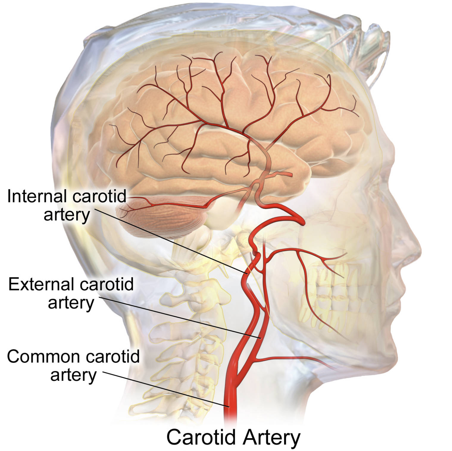
Common carotid artery: This vessel is the main arterial trunk that ascends through the neck to provide blood to the head. The right common carotid arises from the brachiocephalic trunk, while the left originates directly from the aortic arch, making it slightly longer than its right-sided counterpart.
External carotid artery: This major branch begins at the upper border of the thyroid cartilage and travels upward to supply the exterior of the head and the face. It gives rise to eight primary branches, including the facial, maxillary, and superficial temporal arteries, which nourish the muscles of mastication and the scalp.
Internal carotid artery: This artery is responsible for supplying the anterior portion of the brain and the eyes. It ascends to the base of the skull, entering through the carotid canal, where it contributes significantly to the formation of the Circle of Willis to ensure redundant blood flow to cerebral tissues.
The carotid system is meticulously designed to maintain stable blood pressure and oxygen delivery. At the point where the common carotid splits, a specialized area called the carotid sinus contains baroreceptors that monitor systemic blood pressure. Additionally, the carotid body, located in the same vicinity, acts as a chemoreceptor to detect changes in oxygen and carbon dioxide levels in the blood, triggering compensatory respiratory and cardiovascular responses when necessary.
Clinically, the bifurcation of the common carotid artery is a site of significant interest because it is a common location for the development of plaque. The turbulence created by the branching pattern can contribute to the accumulation of fatty deposits, a process known as atherosclerosis. If these plaques become unstable or narrow the vessel significantly, they can lead to transient ischemic attacks or full-scale strokes.
Key features of the carotid system include:
- The division into internal and external branches at the level of the C4 vertebra.
- The lack of branches in the common and internal carotid arteries within the neck.
- The extensive network of the external carotid artery supplying the skin and meninges.
- The role of the internal carotid in providing nearly 80% of the brain’s blood supply.
Physiological Role in Cerebral Hemodynamics
The internal carotid artery is a masterpiece of biological engineering, traveling a complex path to reach the brain. Unlike most arteries, it has a “S-shaped” curve known as the carotid siphon, which helps dampen the pulsatile pressure of the blood before it reaches the delicate tissues of the brain. This mechanism is vital for maintaining steady hemodynamics, protecting the cerebral capillaries from damage caused by high-pressure surges from the heart.
Once inside the cranium, the internal carotid branches into the anterior and middle cerebral arteries. These vessels are responsible for supplying the areas of the brain involved in motor control, sensory perception, and language. Because the brain is highly sensitive to hypoxia, any interruption in this flow, even for a few minutes, can result in permanent neurological deficits.
External Drainage and Facial Supply
While the internal branch looks inward, the external carotid artery focuses on the superficial and deep structures of the head. Its branches are highly interconnected, allowing for excellent collateral circulation in the event of minor injuries to the face or scalp. For example, the facial artery provides the blood needed for the complex movements of the facial muscles, while the maxillary artery supplies the deep structures of the jaw and teeth.
Surgically, the external carotid is a landmark for various procedures involving the neck and throat. Because it has multiple accessible branches, it is often used in reconstructive surgery or as a point of access for interventional radiology. Understanding the specific exit points of these branches allows surgeons to navigate the neck safely while preserving vital blood flow to the skin and sensory organs.
The carotid artery system represents the intersection of structural complexity and functional necessity. From the regulatory receptors at its bifurcation to the delicate branches within the skull, every part of this system is tuned to support the most vital organs of the human body. By maintaining a clear understanding of this anatomy, healthcare professionals can better predict vascular risks and provide effective care for patients with cerebrovascular conditions.

