Understanding the structural intricacies of bacterial pathogens is crucial for modern medicine and microbiology, as it allows researchers to identify disease mechanisms and develop effective treatments. By utilizing advanced imaging techniques like Transmission Electron Microscopy (TEM) and Scanning Electron Microscopy (SEM), researchers can observe everything from internal cell organelles to the surface topography of dangerous bacteria like Staphylococcus aureus. The images provided offer a side-by-side comparison of these two powerful microscopic technologies, highlighting how different methods reveal unique aspects of microbial life.
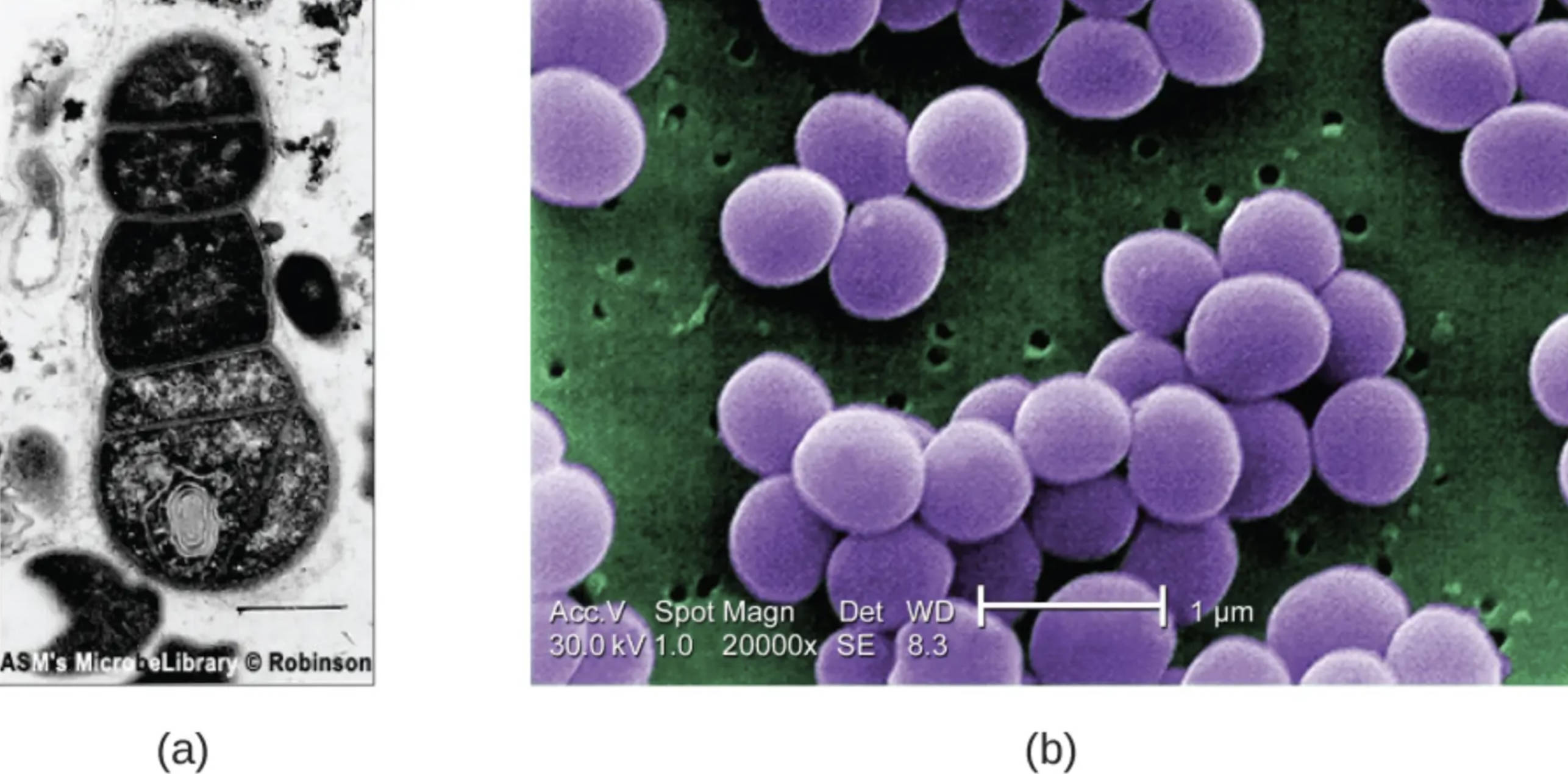
(a): This label indicates a TEM image of bacterial cells embedded within a biofilm, captured in cross-section to reveal the internal complexity of the organism. Because Transmission Electron Microscopy transmits electrons through a thinly sliced specimen, the varying levels of opacity allow scientists to clearly distinguish well-defined internal structures such as the nucleoid region and ribosomes.
(b): This label points to a color-enhanced SEM image of Staphylococcus aureus, showcasing the bacterium’s characteristic spherical shape and clustering habit. Unlike TEM, Scanning Electron Microscopy bounces electrons off the specimen’s surface, rendering a detailed three-dimensional image that highlights the external texture and physical arrangement of the cells.
The Role of Advanced Microscopy in Microbiology
The study of microorganisms has evolved significantly since the invention of the simple light microscope, moving toward high-resolution electron microscopy to visualize the smallest components of life. While light microscopy is useful for general observation, it lacks the resolving power to show minute details of viral particles or the internal organelles of bacteria. Electron microscopy bridges this gap by using beams of electrons instead of light photons, allowing for magnification levels that can reach up to 2 million times. This capability is essential for identifying specific bacterial strains and understanding how they interact with human tissue.
In the context of the provided images, the focus is on two distinct aspects of bacterial analysis: internal composition and surface morphology. The TEM image (a) is particularly valuable for studying the formation of biofilms—protective layers that bacteria secrete to shield themselves from antibiotics and the immune system. By looking inside the cell, researchers can assess the metabolic state of the bacteria and the density of the surrounding matrix. On the other hand, the SEM image (b) provides a topographical map of the pathogen. This 3D perspective is vital for observing cell division, the formation of colonies, and the physical interaction between bacteria and medical devices, such as catheters or implants.
The distinction between these imaging techniques is vital for accurate diagnosis and research. While they both utilize electron beams, their applications differ based on what the pathologist or microbiologist needs to see. To summarize the specific utility of these technologies:
- Transmission Electron Microscopy (TEM): Best for viewing internal structures, cross-sections of cells, and viral internal components; provides a 2D image.
- Scanning Electron Microscopy (SEM): Best for viewing surface textures, overall cell shape, and colony organization; provides a 3D image.
- Sample Preparation: TEM requires very thin slicing of samples, whereas SEM requires coating the sample in a conductive metal (like gold) to reflect electrons.
- Medical Application: TEM is often used in renal pathology and virology, while SEM is frequently used to study infection sites and biofilm development on surfaces.
Clinical Significance of Staphylococcus aureus
The bacterium depicted in image (b) is Staphylococcus aureus, a gram-positive, round-shaped bacterium that is a member of the Firmicutes, and it is frequently found in the upper respiratory tract and on the skin. While it is a common part of the human microbiota, S. aureus is a facultative anaerobe that can become a severe opportunistic pathogen. It is a leading cause of skin and soft tissue infections such as abscesses, furuncles, and cellulitis. However, its medical significance extends far beyond surface infections. If the bacteria enter the bloodstream, they can cause invasive and life-threatening conditions, including pneumonia, osteomyelitis (bone infection), endocarditis (infection of the heart valves), and sepsis.
One of the most pressing challenges in treating S. aureus infections is the emergence of antibiotic-resistant strains, most notably Methicillin-resistant Staphylococcus aureus (MRSA). MRSA is resistant to beta-lactam antibiotics, which include penicillins and cephalosporins, making standard treatments ineffective. This resistance compels healthcare providers to rely on stronger, more toxic antibiotics like vancomycin. The ability of S. aureus to form biofilms, as hinted at in image (a), further complicates treatment. A biofilm is a slimy, glue-like substance that anchors the bacteria to tissues or medical implants, creating a physical barrier that prevents antibiotics and immune cells from reaching the bacteria.
From a physiological standpoint, S. aureus is distinguished by its “grape-like” cluster arrangement, clearly visible in the SEM image. This clustering is a result of the bacteria dividing in multiple planes. The bacterium produces various virulence factors, including enzymes that clot blood (coagulase) to hide from the immune system and toxins that destroy white blood cells (leukocidins). Understanding these physical and chemical defense mechanisms through microscopy helps researchers design new drugs that can penetrate biofilms or disrupt the cell wall of the bacteria.
Conclusion
The visualization of microbial structures through TEM and SEM is more than just a technical achievement; it is a cornerstone of infectious disease pathology. By comparing the internal cross-sections of biofilms with the external 3D structure of pathogens like Staphylococcus aureus, medical science gains a comprehensive view of how these organisms survive, reproduce, and resist treatment. These imaging tools remain indispensable in the ongoing battle against antibiotic resistance, allowing scientists to see the enemy clearly and develop targeted strategies to protect human health.

