This diagram illustrates the critical technique for adult cardiopulmonary resuscitation (CPR), specifically focusing on the correct hand placement for chest compressions. CPR is a life-saving emergency procedure performed when the heart stops beating, aiming to maintain blood flow to the brain and other vital organs until professional medical help arrives. Understanding the anatomical landmarks and the mechanics of effective chest compressions, as depicted here, is paramount for anyone learning this essential skill. This guide will delve into the proper methodology to maximize the chances of a positive outcome during a cardiac arrest event.
T1-T12: These numbers represent the thoracic vertebrae, which are the 12 bones of the spinal column located in the chest region. Each vertebra plays a crucial role in forming the posterior wall of the thoracic cavity and articulating with the ribs. They serve as important anatomical landmarks for proper hand placement during CPR.
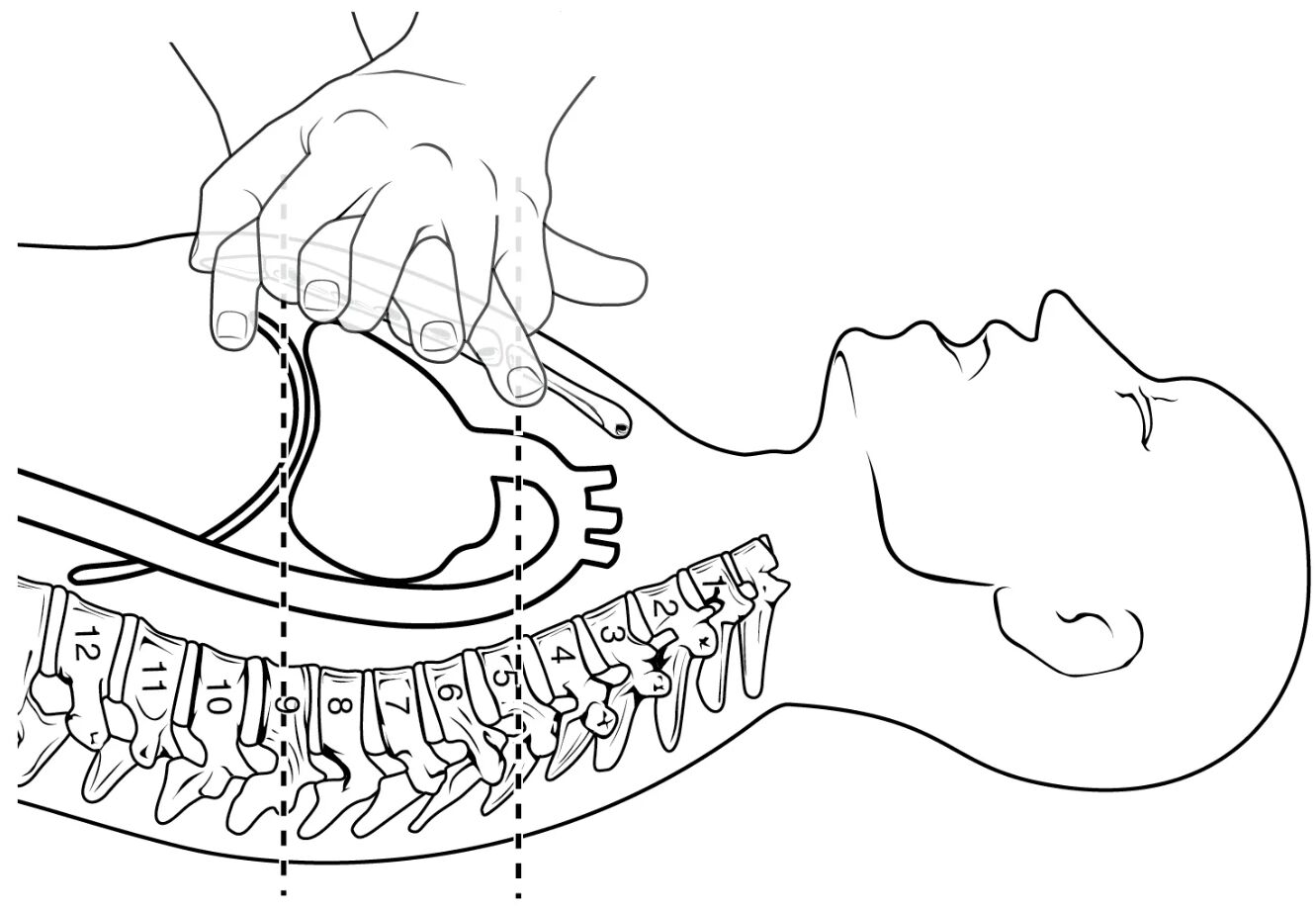
T4: This line indicates the approximate level of the fourth thoracic vertebra. In the context of CPR, the T4 level helps delineate the superior border of the optimal compression area, ensuring compressions are applied over the lower half of the sternum and avoiding the upper chest. Precision in identifying this landmark contributes to effective and safe CPR.
T9: This line marks the approximate level of the ninth thoracic vertebra. The T9 level helps define the inferior border of the optimal compression area, ensuring compressions are performed above the xiphoid process. Proper placement between T4 and T9 is critical to avoid injuries to abdominal organs while maximizing the efficacy of chest compressions.
Introduction to Cardiopulmonary Resuscitation (CPR)
Cardiopulmonary Resuscitation (CPR) is an emergency procedure that can save a person’s life if their breathing or heart stops. This critical intervention is performed when someone experiences cardiac arrest, a sudden and unexpected loss of heart function, breathing, and consciousness. The primary goal of CPR is to maintain a minimal but essential flow of oxygenated blood to the brain and other vital organs until advanced medical care can be provided. Without CPR, brain damage can occur within minutes, and death can follow swiftly. The diagram meticulously illustrates the correct hand placement and body mechanics for adult chest compressions, a cornerstone of effective CPR.
The immediate application of high-quality CPR significantly increases the chances of survival for individuals experiencing cardiac arrest. It essentially acts as an artificial pump, manually compressing the heart between the sternum and the spine to circulate blood. This sustained circulation, even if minimal, helps to preserve brain function and keep other organs viable until defibrillation or other life-saving treatments can be administered. Understanding and correctly executing the technique, as detailed by the anatomical landmarks shown in the image, is fundamental for anyone who might find themselves in a position to save a life.
CPR involves two main components: chest compressions and rescue breaths. While the image focuses on chest compressions, both are crucial for optimizing patient outcomes. The sequence and emphasis on these components can vary slightly depending on the training guidelines (e.g., lay rescuer vs. healthcare professional), but chest compressions remain the priority. This article will specifically detail the correct technique for chest compressions to ensure efficacy and minimize injury.
Key situations where CPR is urgently needed include:
- Sudden Cardiac Arrest: Often caused by an electrical disturbance in the heart.
- Drowning: When water enters the lungs, inhibiting breathing.
- Choking: Obstruction of the airway preventing breathing.
- Electrocution: High voltage shock can disrupt heart rhythm.
- Severe Trauma: Injuries that lead to cessation of breathing or heartbeat.
- Drug Overdose: Certain substances can depress respiratory and cardiac function.
Prompt recognition and initiation of CPR in these scenarios are vital.
The Physiology Behind Chest Compressions
When the heart stops beating, the body’s circulation ceases, leading to a rapid depletion of oxygen in the brain and other critical organs. Chest compressions aim to artificially mimic the pumping action of the heart. By rhythmically applying pressure to the sternum, the heart is squeezed between the sternum and the vertebral column. This mechanical compression increases intrathoracic pressure, which in turn forces blood out of the heart chambers and into the major arteries, propelling it towards the brain, lungs, and other organs. When the pressure is released, the chest recoils, allowing the heart chambers to refill with blood from the venous system.
Effective chest compressions must be performed at an adequate depth and rate. For adults, the recommended depth is at least 2 inches (5 cm) but no more than 2.4 inches (6 cm), and the rate should be between 100 to 120 compressions per minute. Equally important is allowing for complete chest recoil between compressions; this enables the heart to fully refill with blood, ensuring maximum blood flow with each subsequent compression. Without proper recoil, the effectiveness of the compressions is significantly reduced, as less blood is ejected from the heart. Continuous, high-quality chest compressions are the most crucial component of CPR, as they are directly responsible for maintaining vital blood flow.
Proper Hand Placement and Technique
The diagram explicitly details the correct hand placement on the sternum, a critical factor for effective and safe chest compressions. The hands should be positioned over the lower half of the sternum, specifically between the approximate levels of the T4 and T9 thoracic vertebrae. This placement avoids compressing the xiphoid process (the small cartilaginous extension at the lower part of the sternum), which can increase the risk of internal injury, such as liver laceration. To achieve this, rescuers should place the heel of one hand in the center of the chest, usually on the lower half of the sternum, and then place the heel of the other hand on top of the first, interlocking the fingers. The fingers should be kept off the chest wall to ensure that pressure is applied directly through the heel of the hand.
Once the hands are correctly positioned, the rescuer should ensure their arms are straight, and their shoulders are directly over their hands. This alignment allows the rescuer to use their body weight to provide compressions, rather than just arm strength, which helps to achieve the correct depth and reduces fatigue. Compressions should be delivered smoothly and rhythmically, avoiding jerky movements. It is essential to minimize interruptions in chest compressions, as every pause reduces blood flow to the brain. The goal is to maintain continuous, high-quality compressions until the patient recovers, or until advanced medical personnel take over.
The Importance of High-Quality CPR
High-quality CPR is a cornerstone of emergency medical care and significantly impacts the survival rates of individuals experiencing cardiac arrest. The efficacy of CPR is maximized when compressions are performed correctly: at the appropriate depth, rate, and with full chest recoil, while minimizing interruptions. Beyond the technical aspects of hand placement and compression mechanics, proper training and regular refresher courses are vital for anyone who might need to perform CPR. Automated external defibrillators (AEDs) are also crucial adjuncts to CPR, as defibrillation can restart the heart in cases of certain cardiac arrhythmias. The combination of early, high-quality CPR and rapid defibrillation forms the “chain of survival,” drastically improving outcomes for victims of sudden cardiac arrest. Empowering more individuals with the knowledge and skills to perform CPR effectively is a public health imperative.

