This article delves into the critical implications of an Atrial Septal Defect (ASD) when accompanied by cardiomegaly, as revealed by an abnormal chest X-ray. It explains how a hole in the heart’s septum leads to increased blood flow to the lungs and enlargement of the heart chambers, impacting overall cardiac function. Understand the visual evidence of these cardiac changes and their significance in clinical diagnosis.
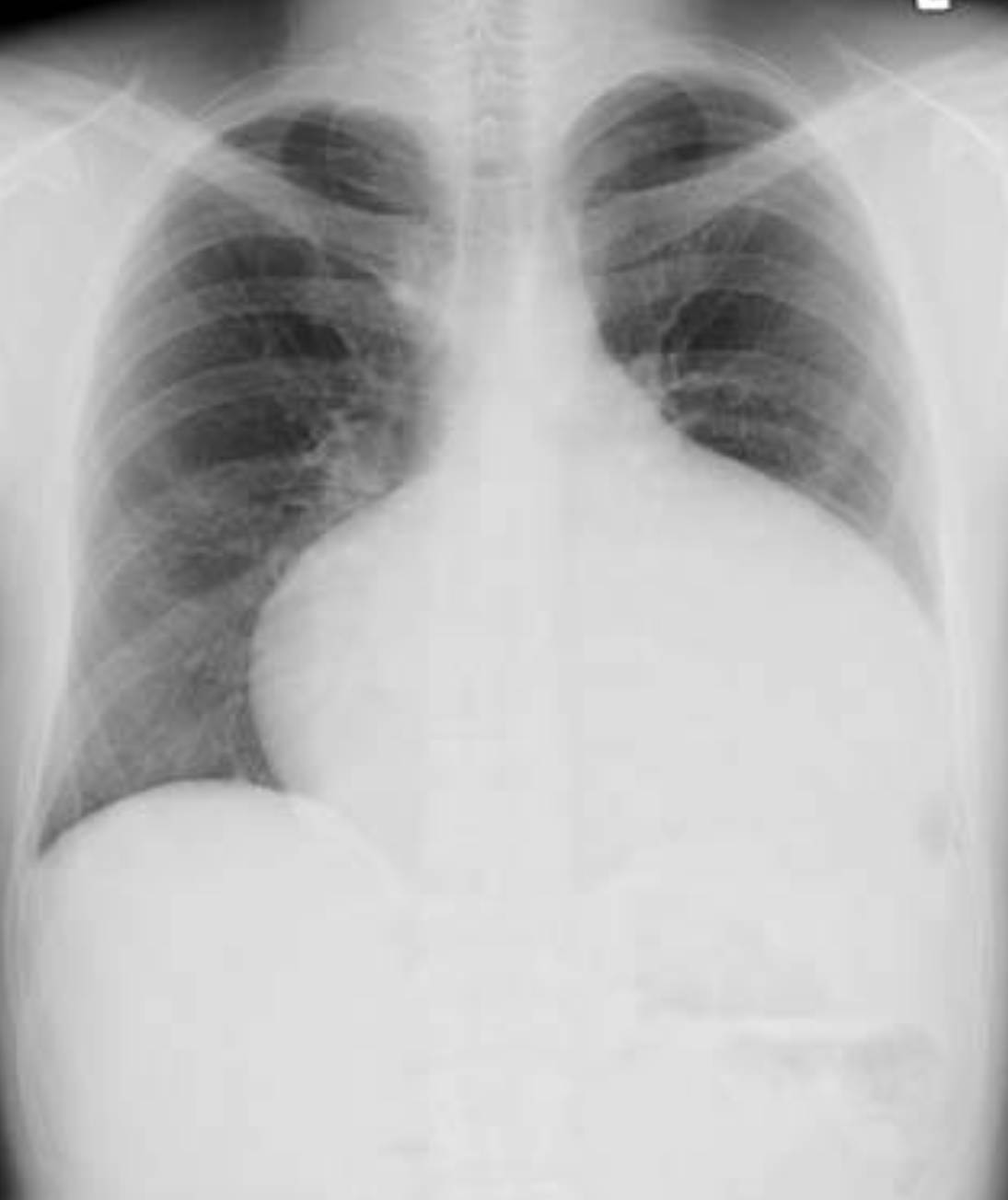
An abnormal chest X-ray in Atrial Septal Defect (ASD) cardiomegaly provides crucial visual evidence of the cardiac changes associated with this congenital heart condition. In a typical chest X-ray, the heart size and lung fields are assessed. When an ASD is present, it often leads to specific patterns that indicate increased blood flow to the lungs and an enlarged heart, primarily affecting the right-sided chambers. These radiographic findings are invaluable for initial diagnosis and monitoring the progression of the disease.
Atrial Septal Defect (ASD) is a congenital heart condition characterized by a hole in the interatrial septum, the wall separating the heart’s two upper chambers (atria). This defect allows oxygen-rich blood from the left atrium to flow into the right atrium, a phenomenon known as a left-to-right shunt. While small ASDs may be asymptomatic and close spontaneously, larger defects can lead to significant physiological consequences due to the increased volume of blood flowing into the right side of the heart and subsequently to the lungs. Over time, this chronic volume overload can cause the heart chambers to enlarge, a condition known as cardiomegaly.
The chest X-ray presented here vividly demonstrates the effects of a significant ASD with associated cardiomegaly. In a healthy individual, the heart silhouette would occupy approximately one-third to one-half of the thoracic cavity width. However, this image clearly shows a markedly enlarged cardiac shadow, indicating an increase in the overall size of the heart. This enlargement is typically due to the dilatation of the right atrium and right ventricle, which work harder to accommodate the extra blood volume shunted from the left side of the heart. The radiographic findings are an important diagnostic clue, prompting further investigation with more definitive cardiac imaging techniques.
The consequences of an uncorrected ASD, especially when accompanied by significant shunting and cardiomegaly, can be profound. The increased blood flow to the lungs can lead to pulmonary hypertension, while the chronic workload on the right side of the heart can eventually result in right-sided heart failure.
- Left-to-right shunt: Oxygenated blood from the left atrium flows to the right atrium.
- Volume overload: Increased blood volume in the right atrium and right ventricle.
- Pulmonary plethora: Increased blood flow to the lungs, visible as engorged pulmonary vessels.
- Cardiomegaly: Enlargement of the heart, particularly the right-sided chambers.
These physiological changes are often detectable on a standard chest X-ray, making it a valuable tool in the initial assessment of suspected congenital heart disease.
The Mechanisms Behind ASD and Cardiomegaly
The presence of an Atrial Septal Defect fundamentally alters the normal blood flow dynamics within the heart. Due to the slightly higher pressure in the left atrium compared to the right atrium, oxygenated blood preferentially flows through the defect from the left atrium to the right atrium. This is termed a left-to-right shunt. This shunted blood then mixes with the deoxygenated blood in the right atrium, increasing the total volume of blood that the right side of the heart must pump.
This chronic volume overload directly impacts the right atrium and the right ventricle. Both chambers must work harder to propel the increased blood volume towards the pulmonary artery and into the lungs. Over an extended period, this sustained increased workload causes the walls of these chambers to stretch and thicken, leading to their enlargement. This is the physiological basis of the cardiomegaly observed on the chest X-ray. Concurrently, the excess blood flow into the pulmonary circulation can cause the pulmonary arteries to dilate and become more prominent, a finding sometimes referred to as “pulmonary plethora” or increased vascular markings, which can also be discernible on the X-ray. Without intervention, this progressive enlargement and increased pulmonary blood flow can eventually lead to irreversible changes in the pulmonary vasculature.
Clinical Implications and Diagnosis of ASD
The clinical implications of an ASD with cardiomegaly vary depending on the size of the defect and the magnitude of the left-to-right shunt. Small ASDs may be entirely asymptomatic, while larger ones can lead to symptoms that become more apparent in adulthood. Common symptoms include shortness of breath, especially during exertion, fatigue, heart palpitations (due to atrial arrhythmias like atrial fibrillation, which can develop secondary to right atrial enlargement), and recurrent respiratory infections in children due to increased pulmonary blood flow. In advanced cases, if pulmonary hypertension becomes severe, it can lead to Eisenmenger syndrome, a rare but serious condition where the shunt reverses from right-to-left, causing cyanosis.
Diagnosis often begins with a physical examination where a characteristic heart murmur may be heard due to the increased blood flow across the pulmonary valve. A chest X-ray is often one of the first diagnostic tools used, providing a general overview of the heart size and lung fields, as demonstrated by the image. The findings of cardiomegaly (particularly right atrial and right ventricular enlargement) and increased pulmonary vascular markings are strong indicators of a significant left-to-right shunt. However, the definitive diagnosis of ASD is typically made with an echocardiogram, which provides detailed images of the heart’s structure, allows direct visualization of the defect, and quantifies the blood flow across the shunt. Other tests, such as an electrocardiogram (ECG) and cardiac MRI, may also be used to assess the heart’s electrical activity and provide further anatomical detail.
Management and Prognosis
The management of Atrial Septal Defect, especially when associated with cardiomegaly, depends on the size of the defect, the extent of shunting, the presence of symptoms, and the development of complications. Small, asymptomatic ASDs that do not cause significant hemodynamic changes may simply require regular monitoring. However, for larger defects causing significant shunting, symptoms, or signs of right-sided heart enlargement and pulmonary overload, intervention is usually recommended to prevent long-term complications.
The primary goal of intervention is to close the defect. This can be achieved through two main approaches:
- Catheter-Based Closure: For secundum ASDs (the most common type), a minimally invasive procedure can be performed where a device (such as a septal occluder) is delivered through a catheter inserted into a blood vessel and deployed to seal the hole.
- Surgical Closure: Open-heart surgery may be necessary for larger or more complex ASDs (e.g., primum or sinus venosus defects) or when catheter closure is not anatomically feasible. During surgery, the defect is either directly stitched closed or patched with synthetic material or the patient’s own pericardial tissue.
After successful closure, the heart typically remodels over time, with the right-sided chambers often reducing in size, and pulmonary blood flow normalizing. The long-term prognosis for individuals with ASD is excellent, especially when the defect is closed before the development of irreversible pulmonary hypertension. Lifelong follow-up with a cardiologist is recommended to monitor for any residual issues or late complications.
The abnormal chest X-ray in Atrial Septal Defect with cardiomegaly provides a crucial initial window into the significant cardiovascular changes induced by this congenital condition. While an ASD represents a structural anomaly, its physiological consequences, particularly the volume overload on the right heart and lungs, can lead to serious health issues if unaddressed. Prompt diagnosis through imaging and appropriate intervention, whether catheter-based or surgical, are vital to restoring normal cardiac function and ensuring a healthy future for affected individuals.

