Delve into the dynamic process of blood circulation through the human heart with this clear and engaging animated guide, showcasing the movement of blood through its four chambers. This visual explanation simplifies the complex pathways of both deoxygenated and oxygenated blood, crucial for sustaining life. Understanding the direction and purpose of blood flow within the heart is fundamental to comprehending cardiovascular health and disease.
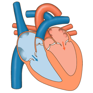
ANIMATED GUIDE CHAMBERS: This label indicates that the image serves as a visual aid to understand the continuous, rhythmic movement of blood through the heart’s compartments. The dynamic representation helps in visualizing the four-chambered pump at work.
Right Atrium: This upper-right chamber of the heart receives deoxygenated blood returning from the body through the superior and inferior vena cava (implied by the large blue vessel entering it). It then contracts to push this blood into the right ventricle.
Left Atrium: Situated in the upper-left, this chamber receives oxygenated blood from the lungs via the pulmonary veins (indicated by the blue vessel with red arrows entering it). It acts as a collecting chamber before transferring blood to the left ventricle.
Left Ventricle: This powerful lower-left chamber is responsible for pumping oxygenated blood into the aorta and subsequently to the rest of the body (indicated by the red vessel exiting it with red arrows). Its thick muscular walls generate the high pressure needed for systemic circulation.
Aorta: The aorta is the largest artery in the body, originating from the left ventricle. It arches over the heart and distributes oxygenated blood to all parts of the systemic circulation, as shown by the red vessel with red arrows leading away.
Pulmonary Artery: This major artery carries deoxygenated blood from the right ventricle to the lungs (indicated by the blue vessel with blue arrows leading towards the lungs). It is unique among arteries for transporting deoxygenated blood.
Right Ventricle: The lower-right chamber of the heart, the right ventricle, pumps deoxygenated blood into the pulmonary artery (as shown by the blue arrows indicating flow out of this chamber). This blood then travels to the lungs for oxygenation.
Aoric Venticle (likely “Aortic Ventricle” or referring to the outflow from the Left Ventricle): While “Aoric Venticle” appears to be a mislabeling or typo, in context, it refers to the left ventricle, which is responsible for ejecting blood into the aorta. This chamber’s strong contraction is essential for systemic blood delivery.
The human heart is an extraordinary organ, functioning as a dual pump to ensure the continuous circulation of blood, delivering oxygen and nutrients to every cell while removing waste products. This diagram elegantly illustrates the distinct pathways of blood flow: deoxygenated blood (shown in blue arrows) from the body enters the right side of the heart, while oxygenated blood (shown in red arrows) from the lungs enters the left side. This crucial separation prevents the mixing of blood with different oxygenation levels, optimizing the efficiency of both pulmonary and systemic circulation.
The journey of deoxygenated blood begins as it enters the Right Atrium, then moves into the Right Ventricle, which subsequently pumps it to the lungs via the Pulmonary Artery for oxygenation. Once oxygenated, the blood returns from the lungs to the Left Atrium, then flows into the powerful Left Ventricle. From there, it is forcefully ejected into the Aorta, which distributes this oxygen-rich blood to the entire body. This sequential movement is tightly regulated by the heart’s valves, which ensure unidirectional flow and prevent backflow.
Understanding this precise pathway of blood flow through the heart is not only fundamental to cardiac physiology but also crucial for recognizing the impact of various cardiovascular conditions. For instance, congenital heart defects, such as a ventricular septal defect (a hole in the wall separating the ventricles), can lead to the mixing of oxygenated and deoxygenated blood, reducing the efficiency of oxygen delivery to the body. Similarly, conditions like pulmonary hypertension can increase the workload on the right ventricle, impacting its ability to pump blood to the lungs effectively.
- The heart pumps approximately 5 liters of blood per minute at rest.
- The entire blood volume circulates through the body in about one minute.
- The right side of the heart handles deoxygenated blood, forming the pulmonary circuit.
- The left side of the heart handles oxygenated blood, forming the systemic circuit.
This animated guide to blood flow provides an invaluable educational tool for both medical professionals and individuals seeking to comprehend the intricate mechanics of the human heart. A thorough appreciation of these pathways and chambers is essential for understanding how the heart sustains life and for recognizing the importance of maintaining cardiovascular health through lifestyle choices and medical care. The heart’s tireless work is a testament to the marvel of biological engineering.

