Explore the intricate world of early avian development through the dorsal view of a 33-hour chick embryo, revealing the nascent structures of the brain, spinal cord, and somites. This article dissects the foundational anatomical changes occurring within the first two days of incubation, offering insights into the rapid organization of a vertebrate body plan. Discover how the primitive streak, neural folds, and somites orchestrate the initial stages of organogenesis.
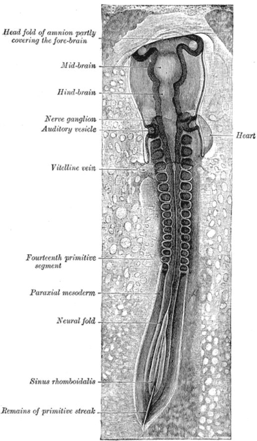
Head fold of amnion partly covering the fore-brain: The amnion is one of the extraembryonic membranes that develops to protect the embryo. The head fold of the amnion is an anterior extension that begins to grow over the developing forebrain, providing a protective covering and contributing to the formation of the amniotic cavity.
Mid-brain: This is the central division of the three primary brain vesicles that develop from the neural tube. The midbrain plays a crucial role in relaying sensory and motor information, and in the adult, it is involved in vision, hearing, and motor control.
Hind-brain: The posterior division of the three primary brain vesicles, the hindbrain will differentiate into structures like the pons, cerebellum, and medulla oblongata in the adult. It is responsible for vital functions such as breathing, heart rate, and coordination of movements.
Nerve ganglion: A nerve ganglion is a cluster of nerve cell bodies located outside the central nervous system. These early ganglia are essential for the development of the peripheral nervous system, transmitting sensory information and motor commands.
Auditory vesicle (Otocyst): This is a primordial structure that develops into the inner ear, including the cochlea and vestibular system. It arises from the ectoderm and is critical for the development of hearing and balance organs.
Heart: At this early stage, the heart is a primitive tube-like structure that has just begun to beat, initiating the circulation of blood. Its formation is one of the earliest and most vital organogenic events, crucial for nutrient and oxygen supply to the rapidly growing embryo.
Vitelline vein: This blood vessel is responsible for draining blood from the yolk sac, which is rich in nutrients, back towards the embryonic heart. The vitelline circulation is crucial for nourishing the developing chick embryo during its early stages.
Fourteenth primitive segment (Somite): Primitive segments, or somites, are blocks of paraxial mesoderm that form segmentally along the anterior-posterior axis of the embryo. Each somite will differentiate into various tissues, including vertebrae, skeletal muscles of the trunk and limbs, and dermis of the skin. The fourteenth segment indicates the extent of axial development at 33 hours.
Paraxial mesoderm: This is a region of mesoderm located alongside the neural tube, which segments to form somites. The paraxial mesoderm is crucial for the development of the axial skeleton, skeletal muscles, and dermis.
Neural fold: These are the lateral edges of the neural plate that elevate and eventually fuse to form the neural tube. The closure of the neural folds is a critical process called neurulation, which forms the precursor to the central nervous system.
Sinus rhomboidalis: This is an unclosed region of the neural tube at the posterior end of the embryo, often seen before the complete closure of the neural folds in the caudal region. It signifies the ongoing process of neural tube formation.
Remains of primitive streak: The primitive streak is a transient structure that forms during gastrulation, through which cells migrate to form the mesoderm and endoderm. Its remains at 33 hours indicate the caudal end of the embryo where gastrulation is still concluding.
The chick embryo, a cornerstone model in developmental biology, offers unparalleled insights into the early stages of vertebrate ontogeny. By 33 hours of incubation, the embryo has undergone profound transformations since fertilization, establishing its fundamental body plan and initiating the formation of major organ systems. A dorsal view at this stage vividly illustrates the progression of neurulation, the segmentation of the paraxial mesoderm, and the nascent circulatory system, all working in concert to shape the future organism. This period is characterized by rapid cellular differentiation and tissue organization, laying the groundwork for complex anatomical structures.
At this critical juncture, the development of the central nervous system is particularly prominent. The neural tube, which is the embryonic precursor to the brain and spinal cord, is largely formed, with distinct regions of the forebrain, midbrain, and hindbrain becoming discernible. These early brain vesicles highlight the cephalic differentiation occurring at the anterior end of the embryo. Simultaneously, the neural folds are still observable in the more caudal regions, signifying the ongoing process of neural tube closure, which is essential for proper nervous system development. The emergence of nerve ganglia further underscores the burgeoning complexity of the nervous system.
Beyond the neural axis, the segmented blocks of paraxial mesoderm, known as somites, are clearly visible flanking the neural tube. These somites are fundamental to the metameric organization of the vertebrate body, as each will give rise to specific vertebrae, muscles, and dermal components. The presence of the heart, already beginning to beat, and the prominent vitelline vein, draining nutrients from the yolk sac, signify the rapid establishment of a functional circulatory system crucial for supporting the embryo’s escalating metabolic demands. The synergy between these developing systems ensures the continued growth and differentiation of the embryo.
Key developmental landmarks at 33 hours include:
- Neural tube formation: Differentiation into brain regions.
- Somite development: Segmental organization of the body.
- Early cardiovascular system: Heartbeat and vitelline circulation.
- Amnion development: Protective membrane formation.
- Remains of primitive streak: Indicating ongoing gastrulation processes.
In conclusion, the 33-hour chick embryo, observed from its dorsal aspect, presents a dynamic tableau of early vertebrate development. The synchronized formation of the nervous system, the segmentation of the mesoderm into somites, and the initiation of cardiovascular function collectively demonstrate the remarkable precision and efficiency of embryogenesis. This detailed anatomical snapshot is invaluable for understanding the foundational processes that underpin the development of all vertebrates, highlighting the intricate biological orchestration that transforms a simple blastoderm into a complex, multi-organism.

