The kidneys are vital organs of the urinary system, playing a critical role in filtering waste products from the blood and maintaining overall body homeostasis. This article provides a comprehensive overview of the kidneys anatomical location, highlighting their protected position within the posterior abdominal wall, shielded by the rib cage and surrounded by adipose tissue. Understanding their precise placement is crucial for comprehending their function and vulnerability to injury.
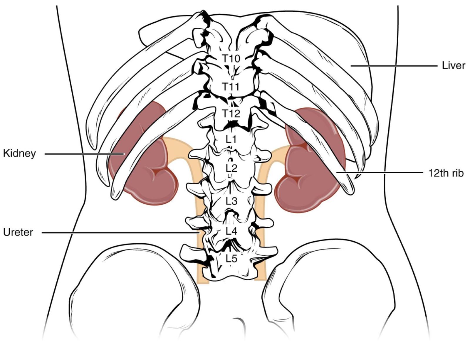
Kidney: These bean-shaped organs are the primary components of the urinary system, responsible for filtering blood to produce urine. They regulate fluid balance, electrolyte levels, and blood pressure, essential for maintaining health.
Ureter: These narrow tubes transport urine from the kidneys to the bladder. Peristaltic contractions propel urine downwards, ensuring a continuous flow and preventing backflow.
Liver: The liver is the largest internal organ, located in the upper right quadrant of the abdomen. It performs a multitude of metabolic functions, including detoxification, protein synthesis, and bile production.
12th rib: This is the lowest pair of ribs, often referred to as a floating rib as it does not directly attach to the sternum. It provides significant protection to the superior pole of the kidney, shielding it from external trauma.
The human kidneys are remarkable organs, tirelessly working to filter approximately 180 liters of blood plasma daily, producing about 1-2 liters of urine. Their precise anatomical positioning is a testament to their importance, strategically placed to receive a degree of natural protection from surrounding skeletal structures. Located retroperitoneally, meaning behind the peritoneum, in the posterior abdominal wall, they reside on either side of the vertebral column. This image offers a posterior view, clearly illustrating their relationship with the lower thoracic and lumbar vertebrae, as well as the ribs.
The kidneys’ placement is not arbitrary; it minimizes their exposure to external forces, crucial for organs so vital to survival.
- Protection by Ribs: The superior poles of both kidneys are partially shielded by the 11th and 12th ribs. The right kidney is typically slightly lower than the left due to the substantial space occupied by the liver above it.
- Adipose Capsule: Beyond the bony protection, each kidney is encased in a thick layer of adipose tissue, known as the perinephric fat or renal fat capsule. This fatty layer provides additional cushioning against physical trauma and helps to hold the kidneys in their proper anatomical position.
This protective arrangement underscores the significance of the kidneys. Their primary function involves filtering waste products, excess salts, and water from the blood, forming urine. This filtration process is carried out by millions of tiny functional units called nephrons within each kidney. Beyond waste removal, the kidneys play a pivotal role in maintaining the body’s acid-base balance, regulating blood pressure through the renin-angiotensin-aldosterone system, and stimulating red blood cell production via erythropoietin. They also convert vitamin D to its active form, which is essential for calcium absorption and bone health.
Given their crucial roles, any damage to the kidneys, whether from trauma or disease, can have profound systemic effects. Conditions like chronic kidney disease (CKD) can impair these functions, leading to the accumulation of toxins, fluid retention, and electrolyte imbalances. In severe cases, kidney failure necessitates dialysis or transplantation. Understanding the anatomical location, including the protection offered by the ribs and surrounding fat, is important not only for surgical approaches but also for recognizing the potential impact of blunt force trauma to the lower back. For instance, a forceful blow to the flank region can cause significant kidney injury despite their protected position, emphasizing the need for awareness and protective measures in sports and high-risk occupations.

