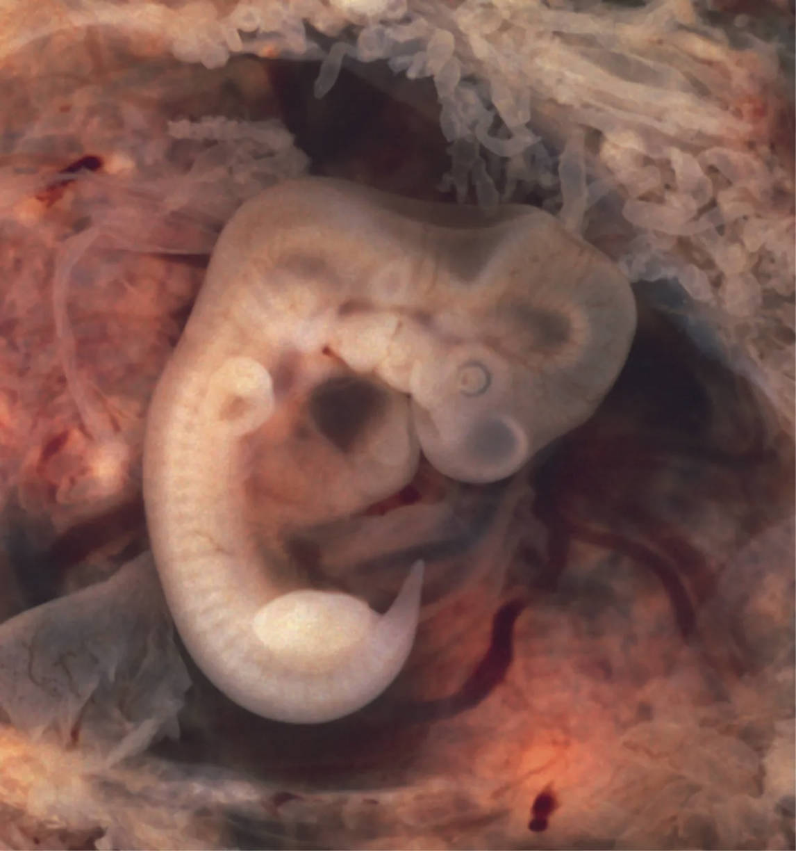
This striking image presents a human embryo at the crucial 7-week mark of development, measuring approximately 10 mm in length. Despite its diminutive size, this stage is characterized by rapid and significant organogenesis, with many foundational structures becoming discernible. The image provides a detailed view of the developing eyes, distinct limb buds, and the prominent tail, illustrating the incredible pace of morphological change during this early gestational period. This particular embryo was derived from an ectopic pregnancy, offering a rare and valuable photographic record of an early developmental stage.
Observable Features of the 7-Week Embryo
While this image does not contain specific labels, several key anatomical features are clearly visible and warrant description.
Developing Eyes: At 7 weeks, the rudimentary structures that will form the eyes are evident as dark, somewhat circular indentations on the side of the head. These optic vesicles are undergoing rapid development, and the lens placode, a thickening of the surface ectoderm, is beginning to invaginate, marking the early stages of lens formation.
Limb Buds: The precursors to the arms and legs are visible as small, paddle-shaped projections from the sides of the embryonic trunk. At this stage, the upper limb buds are typically slightly more developed than the lower limb buds, beginning to show indications of future hand and foot plates.
Tail: A prominent feature of the early embryo, the tail is clearly visible extending caudally. This structure is a normal part of human embryonic development and will regress significantly or be fully incorporated into the developing coccyx by later stages.
Head Region: The cephalic end of the embryo is considerably larger than the trunk, reflecting the rapid growth and differentiation of the brain. The curvature of the head is pronounced due to the flexures forming in the neural tube.
Trunk: The main body of the embryo, from which the limb buds emerge. Within this region, major organ systems are rapidly developing, including the heart (which is already beating), the digestive tract, and early components of the renal system.
Pharyngeal Arches: Although subtle, the developing pharyngeal arches may be faintly discernible in the neck region. These structures are crucial for the formation of the face, neck, and associated structures of the head and throat.
The Rapid Pace of Development at 7 Weeks
The 7-week embryo represents a period of intense and complex development, often referred to as the embryonic period, which spans from fertilization to the end of the 8th week. During this time, the embryo undergoes organogenesis, the process by which all major organ systems begin to form. The heart is actively beating, circulating blood through the rudimentary vascular system. The brain is rapidly expanding, and the neural tube has largely closed, forming the rudimentary central nervous system.
The appearance of limb buds is a significant milestone, as these structures will undergo intricate processes of growth, patterning, and differentiation to form the complex bone, muscle, and joint structures of the upper and lower extremities. Furthermore, the early development of sensory organs, such as the eyes and internal ear, highlights the sophisticated programming guiding embryonic development.
- This specific embryo, originating from an ectopic pregnancy, underscores the critical importance of proper implantation within the uterus for healthy gestation. Ectopic pregnancies, where the embryo implants outside the uterus, are non-viable and can pose significant health risks to the mother.
The detailed visualization of this 7-week embryo offers a powerful testament to the intricate and vulnerable stages of early human life, where every millimeter of growth corresponds to profound biological transformation.
Conclusion
This image of a 7-week embryo provides a fascinating window into the dynamic and rapid changes occurring during early human development. Despite its small size, the presence of developing eyes, limb buds, and a tail signifies the remarkable progress in organogenesis and body plan establishment. While the origin from an ectopic pregnancy highlights a specific medical context, the image primarily serves as an educational tool to appreciate the intricate processes that shape a new life. It underscores the critical period of vulnerability during early gestation when the foundations for all future structures are meticulously laid.

