The ankle joint is a marvel of biomechanical engineering, supporting movement and stability with its complex structure. This article examines the lateral view of the ankle, detailing the bones and ligaments that define its function and highlighting their roles in everyday mobility.
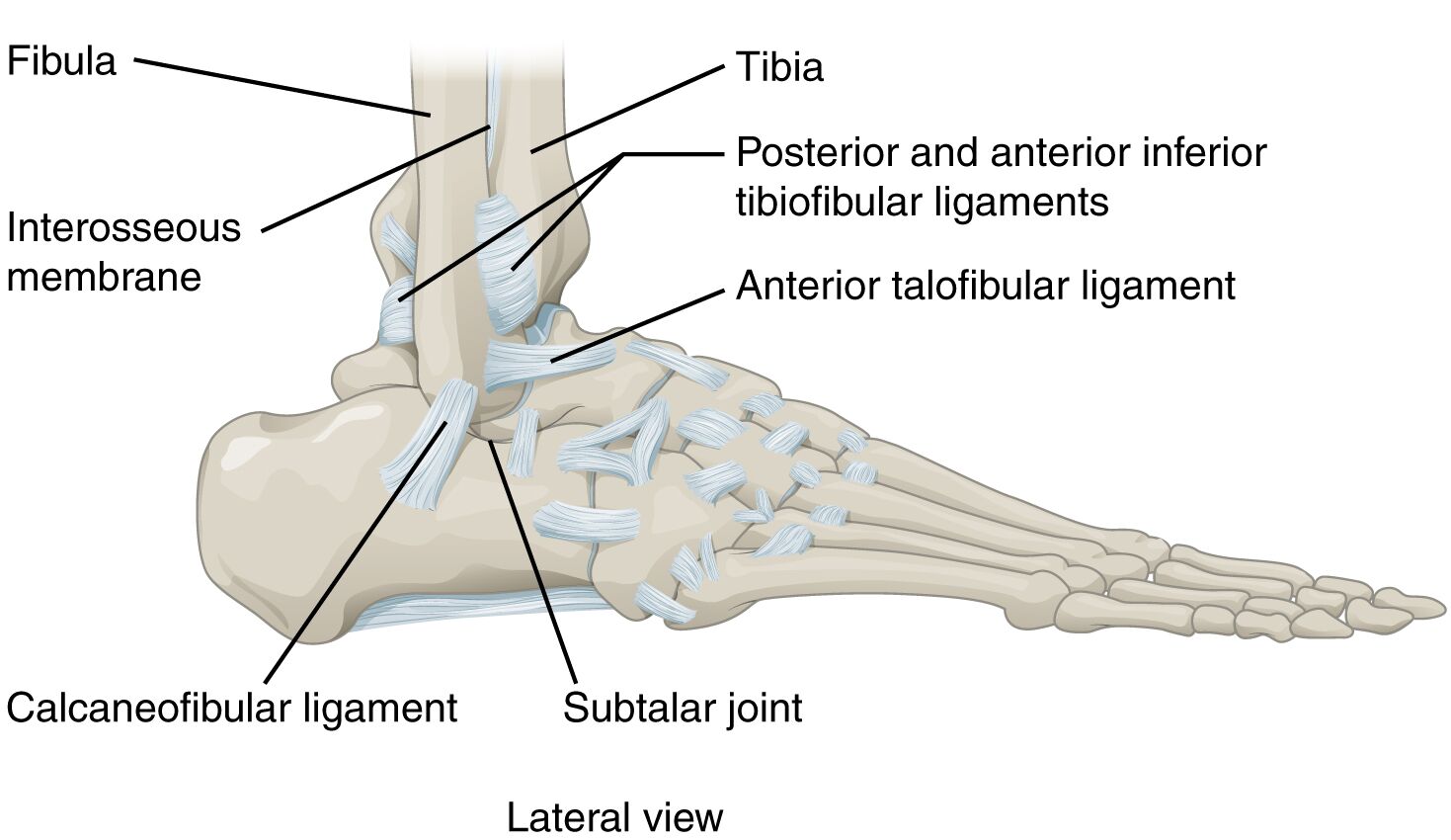
Fibula: The fibula, a slender bone parallel to the tibia, forms the lateral malleolus and provides crucial support to the ankle joint. It plays a key role in stabilizing the lateral aspect, aiding in smooth dorsiflexion and plantar flexion.
Tibia: The tibia, or shinbone, is the larger weight-bearing bone of the lower leg, contributing to the medial and anterior stability of the ankle. It articulates with the talus to form part of the talocrural joint, essential for load distribution.
Posterior and anterior inferior tibiofibular ligaments: These ligaments connect the tibia and fibula at their distal ends, reinforcing the tibiofibular syndesmosis. They ensure proper alignment and stability, preventing excessive separation during movement.
Interosseous membrane: The interosseous membrane is a fibrous structure linking the tibia and fibula along their length. It enhances stability, serves as an attachment for muscles, and assists in force transmission between the bones.
Anterior talofibular ligament: The anterior talofibular ligament runs from the fibula to the talus, offering lateral stability to the ankle. It is frequently injured in ankle sprains due to its susceptibility during inversion movements.
Calcaneofibular ligament: The calcaneofibular ligament extends from the fibula to the calcaneus, providing additional lateral support. It helps resist excessive inversion, contributing to the ankle’s overall resilience.
Subtalar joint: The subtalar joint, located between the talus and calcaneus, enables inversion and eversion of the foot. This joint enhances the ankle’s adaptability, allowing for adjustments to uneven surfaces.
Anatomical Framework and Functionality
The lateral view offers a clear perspective on the ankle’s structural components. The talocrural joint, a uniaxial hinge, permits dorsiflexion and plantar flexion, fundamental for walking.
- The tibia and fibula form a mortise that securely holds the talus, ensuring stability under load.
- The posterior and anterior inferior tibiofibular ligaments maintain the integrity of this mortise, critical for weight-bearing.
- The interosseous membrane distributes forces evenly, reducing stress on individual bones.
- The subtalar joint adds flexibility, enabling the foot to adapt to diverse terrains.
This intricate setup supports the ankle’s role as a dynamic pivot point.
Ligament Roles in Stability
Ligaments are vital for protecting the ankle from injury. The lateral ligaments are particularly significant in maintaining balance.
- The anterior talofibular ligament is a common site of sprains, highlighting its importance in lateral support.
- The calcaneofibular ligament works in tandem, resisting inversion forces that could destabilize the joint.
- The posterior and anterior inferior tibiofibular ligaments ensure the fibula and tibia remain aligned, enhancing overall strength.
- The interosseous membrane provides a continuous link, bolstering the lower leg’s structural unity.
These ligaments absorb shock and maintain alignment, essential for athletic and daily activities.
Clinical Implications and Movement
The ankle’s design supports a range of motions with clinical relevance. The talocrural joint’s hinge action is complemented by the subtalar joint’s side-to-side movements.
- Muscles like the peroneus longus and brevis drive inversion and eversion, relying on ligament stability.
- The anterior talofibular ligament’s vulnerability underscores the need for proper ankle support during sports.
- The subtalar joint’s flexibility aids in balance, adapting the foot to irregular surfaces.
- The calcaneofibular ligament helps prevent chronic instability, a concern in repeated injuries.
This biomechanical harmony is key to understanding ankle function and injury prevention.
The lateral view of the ankle joint reveals a sophisticated interplay of bones and ligaments that ensure mobility and stability. The tibia and fibula provide a robust skeletal framework, while the posterior and anterior inferior tibiofibular ligaments and interosseous membrane reinforce their connection. Ligaments like the anterior talofibular ligament and calcaneofibular ligament offer critical lateral support, with the subtalar joint enhancing adaptability. Appreciating these elements fosters a deeper understanding of the ankle’s role in movement and the importance of protecting it for long-term health.

