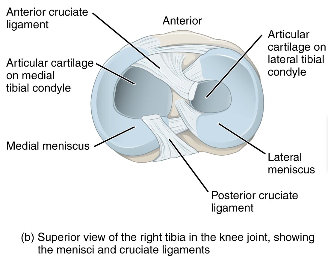
The knee joint stands as the largest and most intricate hinge joint in the human body, pivotal for mobility and weight-bearing activities. This superior view of the right tibia within the knee joint highlights the critical roles of the menisci and cruciate ligaments, offering a window into the structural foundation that supports everyday movements. Delving into this anatomical perspective enhances understanding of the knee’s stability and function, making it an essential study for those interested in human physiology.
Labels Introduction
- Anterior cruciate ligament: The anterior cruciate ligament (ACL) prevents excessive forward movement of the tibia relative to the femur, ensuring knee stability during dynamic activities. It is a common site of injury, particularly in sports involving sudden stops or pivots, often necessitating surgical reconstruction.
- Articular cartilage on medial tibial condyle: This smooth cartilage covers the medial tibial condyle, reducing friction between the tibia and femur during knee movement. It also absorbs shock and distributes load, protecting the underlying bone from wear.
- Articular cartilage on lateral tibial condyle: Located on the lateral tibial condyle, this cartilage facilitates smooth articulation with the femur, minimizing joint wear. It plays a key role in load-bearing and shock absorption, contributing to long-term joint health.
- Medial meniscus: The medial meniscus is a C-shaped fibrocartilage that cushions the medial side of the knee, enhancing stability between the femur and tibia. It also aids in load distribution and can be prone to tears due to its fixed attachment to the joint capsule.
- Lateral meniscus: The lateral meniscus, more circular in shape, acts as a shock absorber on the lateral side of the knee, supporting the femur-tibia interface. Its mobility within the joint helps it adapt to various movements, reducing the risk of injury compared to the medial meniscus.
- Posterior cruciate ligament: The posterior cruciate ligament (PCL) prevents the tibia from sliding backward under the femur, maintaining posterior stability. It is stronger than the ACL and often injured in high-impact trauma, such as dashboard injuries in car accidents.
Anatomical and Physical Introduction
The knee joint’s superior view of the right tibia reveals a sophisticated network of structures that ensure its functionality. The tibial condyles, covered with articular cartilage, provide a smooth surface for the femur to glide over, supported by the medial and lateral menisci. These menisci not only cushion the joint but also enhance congruence between the femoral and tibial surfaces, distributing weight effectively during movement.
The cruciate ligaments, including the anterior and posterior varieties, are vital for rotational and translational stability. The anterior cruciate ligament restricts anterior tibial displacement, while the posterior cruciate ligament counters posterior movement, together forming a dynamic stabilizing system. This intricate balance allows the knee to handle the stresses of walking, running, and jumping, showcasing its biomechanical efficiency.
Beyond ligaments and cartilage, the knee’s design includes collateral ligaments (tibial and fibular) outside the articular capsule, adding lateral support. The synovial fluid within the joint nourishes the cartilage, ensuring its resilience, while the menisci adapt to varying loads. This combination of elements underscores the knee’s role as a resilient yet vulnerable joint, requiring careful attention to maintain its health.
Conclusion
The superior view of the right tibia in the knee joint offers a detailed glimpse into the anatomy that powers human locomotion. The interplay of the menisci, cruciate ligaments, and articular cartilage highlights the knee’s ability to absorb shock and maintain stability under diverse conditions. Gaining insight into these structures fosters a greater appreciation for the knee’s complexity and the importance of protecting it from injury or degeneration.

