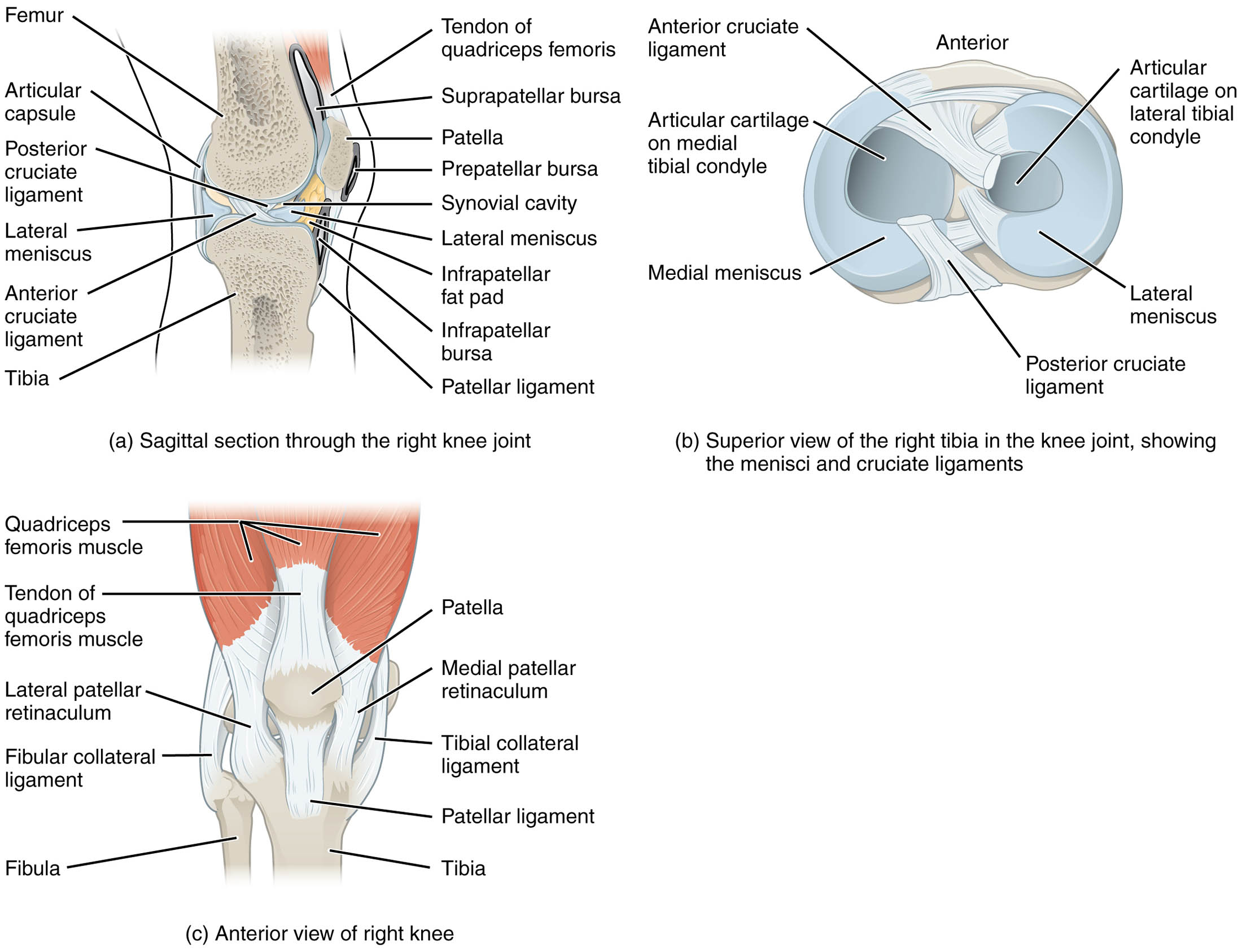
The right knee joint, depicted in sagittal, superior, and anterior views, stands as the largest joint in the body, showcasing a complex interplay of bones, ligaments, and cartilage. This illustration highlights the supporting structures like the cruciate and collateral ligaments, along with the menisci, which provide padding and stability between the femur and tibia. Exploring these components offers valuable insights into how the knee facilitates movement and bears weight in daily activities.
Labeled Parts Explanation
- Femur: This thigh bone forms the upper part of the knee joint, articulating with the tibia to enable flexion and extension. Its condyles play a critical role in weight distribution and joint stability.
- Tibia: The shin bone forms the lower part of the knee joint, providing a stable base for the femur and supporting body weight. It interacts with the menisci to absorb shock during movement.
- Fibular collateral ligament: This ligament on the lateral side connects the femur to the fibula, preventing excessive varus stress. It supports the knee’s side-to-side stability outside the articular capsule.
- Tibial collateral ligament: Located on the medial side, this ligament links the femur to the tibia, resisting valgus stress. It helps maintain knee alignment and is crucial for medial stability.
- Patella: This sesamoid bone sits in front of the knee, protecting the joint and enhancing the leverage of the quadriceps muscle. It glides within the femoral groove during knee flexion.
- Patellar ligament: This strong band connects the patella to the tibia, transmitting forces from the quadriceps to extend the knee. It plays a key role in stabilizing the patella during movement.
- Medial patellar retinaculum: This fibrous structure on the medial side supports the patella, aiding in its tracking within the femoral groove. It helps maintain proper patellar alignment during knee motion.
- Lateral patellar retinaculum: Located on the lateral side, this structure stabilizes the patella and assists in its smooth movement. It works with the medial retinaculum to ensure balanced patellar function.
- Quadriceps femoris muscle: This group of muscles on the anterior thigh extends the knee via the quadriceps tendon. It is essential for activities like standing and walking.
- Tendon of quadriceps femoris muscle: This tendon connects the quadriceps muscle to the patella, facilitating knee extension. It transmits the muscle’s force to the patellar ligament.
- Suprapatellar bursa: This fluid-filled sac above the patella reduces friction between the femur and quadriceps tendon. It cushions the joint during movement and prevents irritation.
- Prepatellar bursa: Located in front of the patella, this bursa minimizes friction between the skin and patella. It protects the knee during kneeling or direct pressure.
- Infrapatellar fat pad: This adipose tissue behind the patellar ligament cushions the joint and fills space within the knee. It also supports the patella’s movement and reduces stress.
- Infrapatellar bursa: This bursa beneath the patella reduces friction between the patellar ligament and tibia. It aids in smooth knee flexion and extension.
- Synovial cavity: This space within the joint contains synovial fluid, lubricating the articular surfaces and nourishing the cartilage. It ensures smooth movement and joint health.
- Articular capsule: This fibrous structure encloses the knee joint, providing stability and containing the synovial cavity. It limits excessive motion while protecting internal components.
- Anterior cruciate ligament: Located inside the capsule, this ligament prevents anterior displacement of the tibia relative to the femur. It is vital for knee stability during rotational movements.
- Posterior cruciate ligament: Also inside the capsule, this ligament prevents posterior displacement of the tibia. It maintains knee integrity, especially during flexion.
- Medial meniscus: This C-shaped cartilage on the medial tibia acts as a shock absorber between the femur and tibia. It enhances joint stability and distributes weight evenly.
- Lateral meniscus: This circular cartilage on the lateral tibia cushions the joint and supports load distribution. It aids in knee stability and reduces wear on articular surfaces.
- Articular cartilage on medial tibial condyle: This smooth layer covers the medial tibia, reducing friction and absorbing shock. It protects the bone during knee movement.
- Articular cartilage on lateral tibial condyle: This cartilage on the lateral tibia minimizes friction and cushions the joint. It ensures smooth interaction with the femoral condyles.
Introduction to Knee Joint Anatomy
The knee joint, the largest in the body, serves as a hinge joint that supports weight and enables flexion, extension, and slight rotation. This illustration presents the right knee in sagittal, superior, and anterior views, highlighting the femur, tibia, and supporting structures like the cruciate ligaments and menisci. A detailed examination of these components reveals how they collaborate to ensure stability and mobility in daily activities.
- Provides a foundational understanding of knee structure.
- Emphasizes the joint’s role in lower body function.
Ligament Support and Joint Stability
Ligaments are essential for maintaining knee integrity, with each playing a specific role. The anterior cruciate ligament prevents the tibia from sliding forward, while the posterior cruciate ligament stops backward movement, both critical for rotational stability. The tibial and fibular collateral ligaments on the sides resist lateral stresses, protecting the joint from side-to-side forces.
- Explains how cruciate ligaments ensure dynamic stability.
- Details the collateral ligaments’ role in lateral support.
Cartilage and Menisci Functions
The menisci and articular cartilage are vital for cushioning and load distribution within the knee. The medial and lateral menisci absorb shock between the femoral and tibial condyles, enhancing joint longevity. The articular cartilage on the tibial condyles reduces friction, ensuring smooth interaction during movement.
- Describes the menisci’s contribution to shock absorption.
- Highlights cartilage’s role in joint lubrication.
Muscle and Tendon Contributions
Muscles and tendons provide the power for knee movement, with the quadriceps femoris muscle extending the joint via its tendon. The patellar ligament transmits this force to the tibia, while the suprapatellar and infrapatellar bursae reduce friction around the patella. The retinacula on both sides stabilize the patella during motion.
- Outlines the quadriceps’ role in knee extension.
- Explains the bursae’s function in friction reduction.
Clinical Relevance and Functional Insights
Understanding knee anatomy is key to addressing issues like ligament tears or meniscus injuries. The synovial cavity’s health supports cartilage nutrition, while the articular capsule limits excessive motion to prevent damage. This knowledge can guide rehabilitation to restore knee function and prevent chronic conditions.
- Offers insight into common knee injuries.
- Suggests the importance of anatomical awareness in treatment.
Conclusion
The right knee joint’s anatomy, as depicted in this illustration, showcases a remarkable balance of bones, ligaments, cartilage, and muscles that enable its function as the body’s largest joint. From the stabilizing cruciate ligaments to the cushioning menisci, each component plays a crucial role in supporting movement and weight. Exploring this anatomy not only deepens your appreciation of the knee’s complexity but also equips you with the insight to maintain its health and address potential issues effectively.

