The posterior view of the right hip joint, with the capsule in place, provides a detailed look at a critical ball-and-socket joint that supports the body’s weight and ensures stability. This illustration highlights the ligaments and bony structures that tighten when standing, pulling the femoral head into the acetabulum, offering insight into their roles in movement and load-bearing. Exploring this image deepens your understanding of the hip’s posterior anatomy and its contribution to lower body functionality.
Labeled Parts Explanation
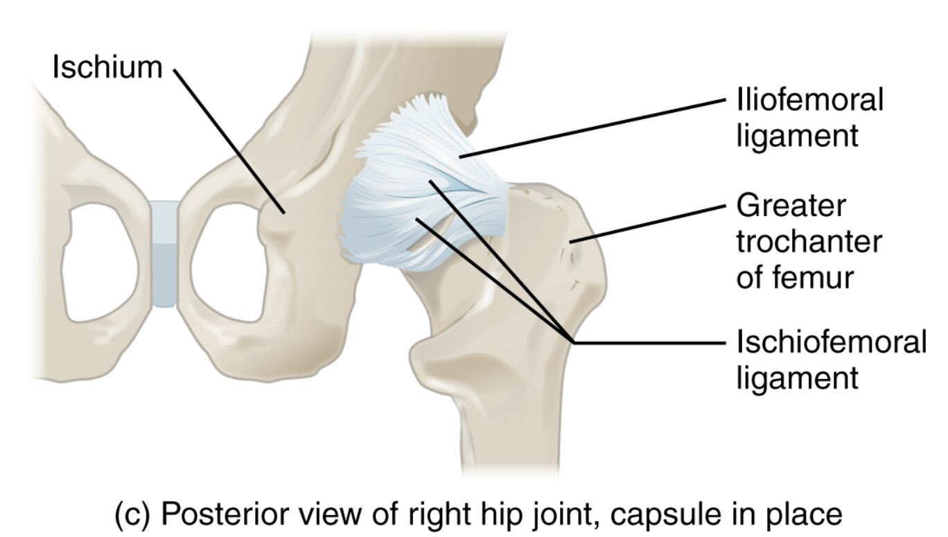
- Ischium: This posterior part of the coxal bone forms the lower boundary of the acetabulum, supporting weight when sitting. It also serves as an attachment site for ligaments and muscles that stabilize the hip.
- Iliofemoral ligament: This robust ligament connects the ilium to the femur, preventing hyperextension and tightening during standing to secure the femoral head. It is one of the strongest ligaments in the body, providing significant joint stability.
- Greater trochanter of femur: This bony prominence on the femur acts as an attachment point for hip abductor muscles like the gluteus maximus. It plays a key role in stabilizing the pelvis during locomotion.
- Ischiofemoral ligament: This ligament links the ischium to the femur, restricting internal rotation and hyperextension of the hip. It complements other ligaments to maintain posterior joint integrity.
Introduction to Hip Joint Anatomy
The hip joint is a foundational structure in the lower body, operating as a ball-and-socket joint that balances stability with a wide range of motions. This posterior view illustration of the right hip joint, with the capsule intact, showcases the ischium and ligaments like the iliofemoral and ischiofemoral, which tighten when standing. A closer look at these elements reveals how they support the femoral head and contribute to everyday movements.
- Offers a comprehensive view of the hip’s posterior structure.
- Emphasizes the joint’s role in weight-bearing and stability.
Ligament Functions in Posterior Stability
Ligaments are crucial for securing the hip from the posterior aspect, each with a specific role. The iliofemoral ligament, known for its strength, prevents overextension and pulls the femoral head into the acetabulum when upright. The ischiofemoral ligament limits internal rotation, ensuring the joint remains aligned during dynamic activities.
- Details how ligaments enhance hip stability when standing.
- Highlights the ischiofemoral ligament’s role in rotation control.
Bony Structures and Their Contributions
The hip’s skeletal framework includes the ischium, a key component of the coxal bone, which supports sitting posture. The greater trochanter of the femur provides a robust attachment for muscles that stabilize the pelvis, essential for walking and standing. These bony landmarks work together to distribute weight and facilitate movement.
- Explains the ischium’s function in weight support.
- Describes the greater trochanter’s role in muscle attachment.
Clinical Relevance and Functional Insights
Understanding the posterior hip anatomy aids in identifying issues like ligament strains or joint instability. The iliofemoral ligament’s strength is vital in preventing posterior dislocations, while the ischiofemoral ligament helps avoid excessive rotation. This knowledge can guide therapeutic approaches to restore hip function and prevent injuries.
- Provides context for potential posterior hip conditions.
- Suggests the importance of anatomical awareness in rehabilitation.
Conclusion
The posterior view of the right hip joint reveals a sophisticated network of ligaments and bones that ensure its stability and support during standing and movement. From the sturdy iliofemoral ligament to the supportive ischium, each structure plays an integral role in maintaining the hip’s functionality. Exploring this anatomy not only fosters a greater appreciation of the hip’s design but also equips you with the insight to address its health and performance effectively.

