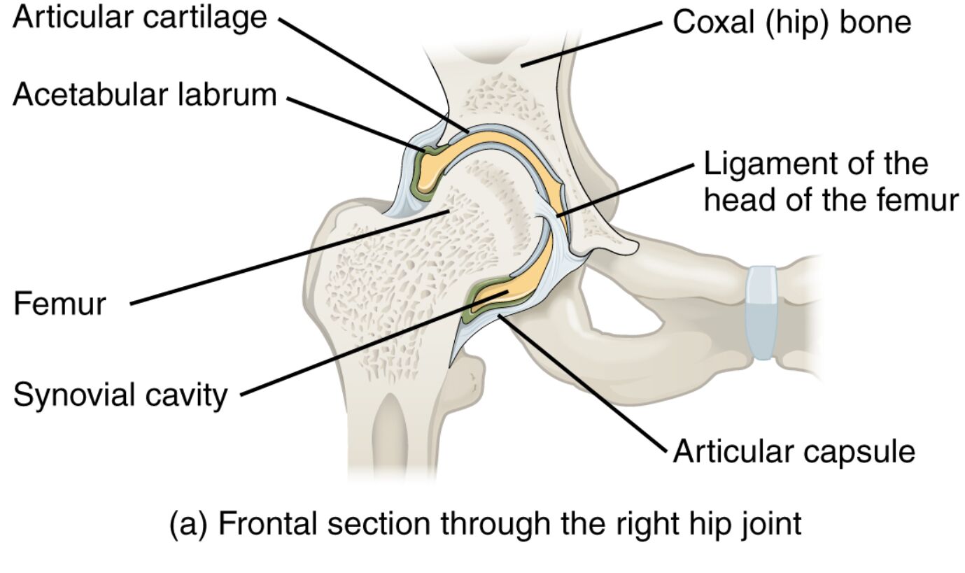
The frontal section of the right hip joint reveals the intricate details of a ball-and-socket joint that supports the body’s weight and enables a wide range of motions. This anatomical illustration highlights the femur, coxal bone, and supporting structures like ligaments and cartilage, offering a clear view of their roles in stability and movement. Exploring this image provides a deeper understanding of how the hip joint contributes to everyday activities and overall lower body mechanics.
Labeled Parts Explanation
- Articular cartilage: This smooth layer covers the femoral head and acetabulum, minimizing friction and absorbing shock during hip movement. It is essential for maintaining joint health by preventing direct bone contact.
- Acetabular labrum: This fibrocartilaginous ring enhances the depth of the acetabulum, securing the femoral head and improving joint stability. It also helps distribute mechanical loads across the hip joint.
- Ligament of the head of the femur: This ligament connects the femoral head to the acetabulum, providing minor stability and housing blood vessels that supply the femoral head. Though not as robust as other ligaments, it supports nutrient delivery to the bone.
- Femur: The thigh bone forms the ball of the hip joint, articulating with the acetabulum to allow multiaxial movement such as flexion and rotation. Its strength is crucial for weight-bearing and locomotion.
- Synovial cavity: This space within the joint contains synovial fluid, which lubricates the articular surfaces and nourishes the cartilage. It ensures smooth motion and reduces wear on joint components.
- Articular capsule: This fibrous envelope surrounds the hip joint, limiting excessive movement while protecting the internal structures. It contains the synovial cavity and contributes to joint integrity.
- Coxal (hip) bone: This pelvic bone forms the socket of the hip joint, known as the acetabulum, and provides a stable base for the femur. It plays a key role in weight distribution and pelvic alignment.
Detailed Article
Introduction to Hip Joint Anatomy
The hip joint serves as a pivotal structure in the lower body, functioning as a ball-and-socket joint that balances stability with mobility. This frontal section illustration showcases the right hip’s key components, including the femur and coxal bone, along with supporting ligaments and cartilage. A thorough understanding of these elements is vital for appreciating how the hip supports activities like walking, running, and standing.
- Offers a detailed look at the hip’s structural design.
- Emphasizes the joint’s role in facilitating diverse movements.
Ligament and Cartilage Roles
Ligaments and cartilage are critical for the hip’s functionality, with each component serving a specific purpose. The acetabular labrum deepens the socket, enhancing stability, while the ligament of the head of the femur provides additional support and vascular supply. The synovial cavity, filled with synovial fluid, ensures the articular cartilage remains lubricated, promoting smooth joint action.
- Explains how the labrum contributes to load distribution.
- Highlights the synovial fluid’s role in cartilage nutrition.
Bone Structure and Joint Mechanics
The hip’s skeletal framework relies on the femur, which forms the ball, and the coxal bone, creating the acetabulum. The articular capsule encases these bones, restricting excessive motion to protect the joint, while the articular cartilage cushions the surfaces. This combination allows the hip to handle significant stress while maintaining flexibility for various movements.
- Details the femur’s contribution to hip strength.
- Describes the coxal bone’s role in pelvic stability.
Clinical Relevance and Functional Insights
Knowledge of hip anatomy is essential for identifying potential issues, such as cartilage degeneration or ligament strain. The articular capsule’s integrity helps prevent dislocations, while the synovial cavity’s health supports long-term joint function. This understanding can guide interventions to maintain or restore hip mobility and strength.
- Provides insight into common hip joint challenges.
- Suggests the importance of anatomical knowledge in clinical practice.
Conclusion
The frontal section of the right hip joint illustrates a sophisticated interplay of bones, ligaments, and cartilage that underpins its remarkable stability and range of motion. From the protective articular capsule to the nourishing synovial cavity, each structure plays a vital role in supporting the body’s daily demands. Exploring this anatomy not only fosters a greater appreciation of the hip’s complexity but also equips you with the insight to address its health effectively.

