Graded potentials play a crucial role in the initial stages of neuronal communication, acting as temporary shifts in the membrane voltage of cells. These changes, influenced by the strength and duration of stimuli, can either depolarize or hyperpolarize the membrane, depending on the specific ion channels activated. This article explores the intricacies of graded potentials, providing a detailed breakdown of the process depicted in the accompanying image, making it an essential resource for understanding how neurons process signals.
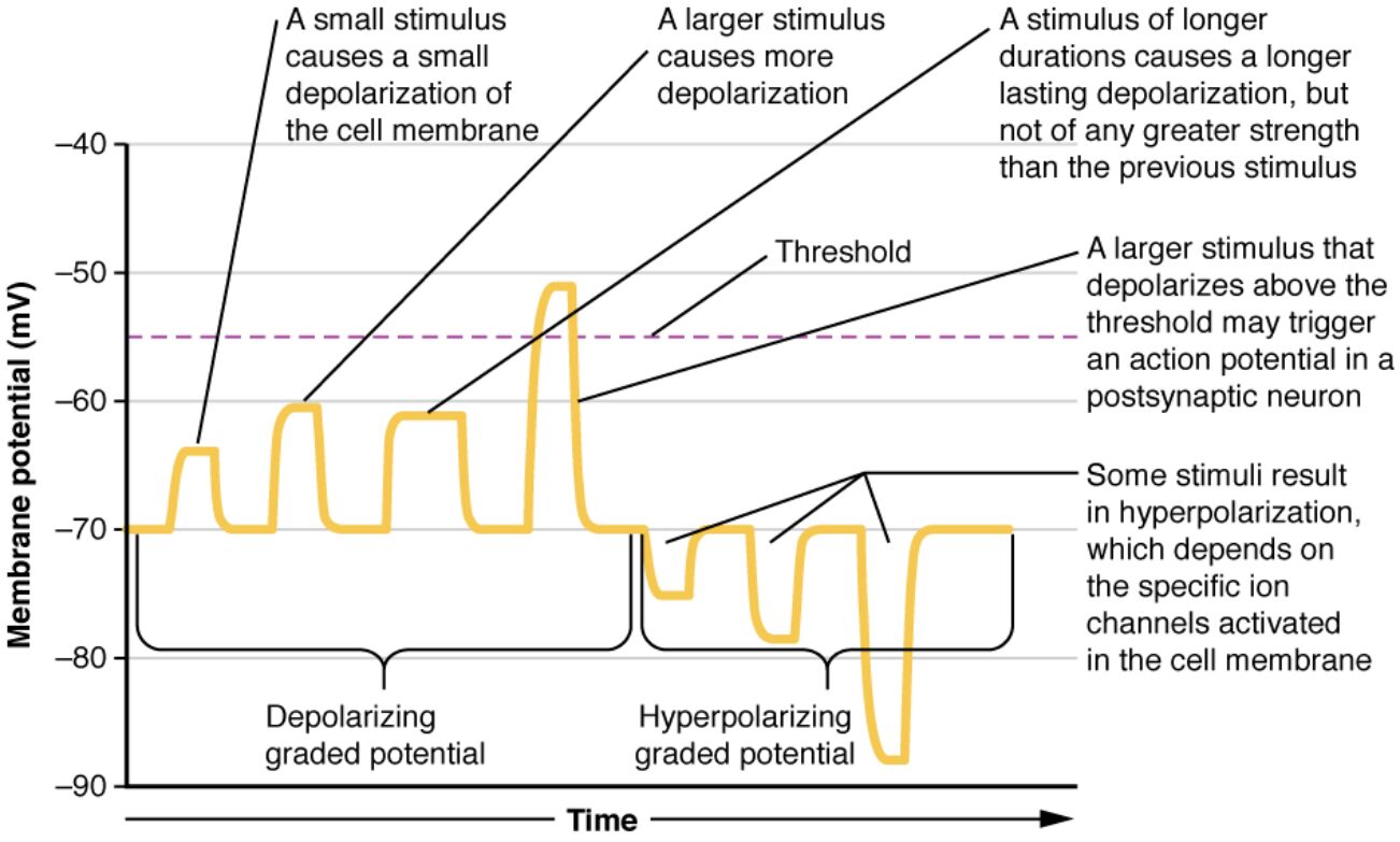
A small stimulus: This label indicates a minimal external trigger that results in a slight depolarization of the cell membrane. The change is localized and proportional to the stimulus strength, not sufficient to reach the threshold for an action potential.
A larger stimulus: This represents a stronger external input causing a more significant depolarization of the membrane. It brings the membrane potential closer to the threshold, though it may still not trigger an action potential unless it exceeds this critical level.
A stimulus of longer durations: This label shows that an extended stimulus can lead to a prolonged depolarization, maintaining the change over time without increasing its amplitude. The duration alone does not enhance the strength beyond the initial larger stimulus effect.
Threshold: This critical membrane potential level, around -50 mV, marks the point where a sufficient depolarization can initiate an action potential. It serves as a boundary that, when crossed, triggers a rapid and significant response in the neuron.
A larger stimulus that depolarizes above the threshold: This indicates a strong enough stimulus that pushes the membrane potential beyond the threshold, potentially initiating an action potential in a postsynaptic neuron. It highlights the all-or-none principle where exceeding this level ensures a full response.
Some stimuli result in hyperpolarization: This label points to stimuli that cause the membrane potential to become more negative, moving away from the threshold. The specific ion channels activated, such as potassium or chloride channels, determine this hyperpolarizing effect.
Depolarizing graded potential: This term describes the positive shift in membrane potential caused by the influx of ions like sodium. It is a graded response that varies with the stimulus intensity and is critical for signal integration.
Hyperpolarizing graded potential: This refers to a negative shift in membrane potential due to the efflux of potassium or influx of chloride ions. It acts to inhibit the neuron, making it harder to reach the threshold for an action potential.
Detailed Explanation of Graded Potentials
Graded potentials are the foundation of how neurons respond to stimuli, initiating the process that may lead to action potentials. These potentials vary in magnitude and duration, directly reflecting the strength and length of the applied stimulus. The image illustrates how different stimuli affect the membrane potential, providing a visual guide to this dynamic process.
- Graded potentials occur in the dendrites and cell body of neurons, where they integrate multiple inputs.
- Depolarization happens when sodium channels open, allowing sodium ions to enter the cell, raising the internal voltage.
- Hyperpolarization results from potassium channels opening or chloride influx, lowering the membrane potential.
- The threshold is a pivotal point; only when depolarization exceeds this level does an action potential propagate.
- These potentials are local and decay with distance, unlike action potentials which are regenerative.
The Role of Ion Channels in Graded Potentials
Ion channels are the gatekeepers of membrane potential changes, playing a vital role in neuronal signaling. Their activation determines whether the cell depolarizes or hyperpolarizes, shaping the response to external stimuli. Understanding their function is key to grasping how neurons process information.
- Specific ion channels, such as voltage-gated sodium channels, facilitate depolarization during a strong stimulus.
- Potassium channels contribute to hyperpolarization by allowing potassium ions to leave the cell.
- Chloride channels can also hyperpolarize the membrane by permitting chloride ion influx.
- The selective opening of these channels depends on the stimulus type and intensity.
- This selective activation ensures precise control over the neuron’s excitability.
Clinical Relevance and Anatomical Context
While graded potentials themselves are not associated with a specific disease, their dysfunction can contribute to neurological conditions. Proper functioning of ion channels and membrane potentials is essential for healthy neuronal communication. This section provides an anatomical and physiological overview to enhance understanding.
- Neurons are anatomically structured with dendrites receiving graded potentials, integrating signals before transmission.
- The cell membrane contains a variety of ion channels, including sodium, potassium, and chloride, which regulate potential changes.
- Physiologically, graded potentials allow neurons to sum excitatory and inhibitory inputs.
- This integration process occurs in the postsynaptic region, influencing whether an action potential is generated.
- Disruptions in ion channel function can lead to altered signaling, potentially affecting neural coordination.
Graded potentials are a fundamental aspect of neuronal physiology, serving as the stepping stone to action potentials that drive communication across the nervous system. The image provides a clear depiction of how varying stimuli influence membrane potential, offering a valuable tool for learning. By mastering these concepts, one can better appreciate the complexity of neural signaling and its importance in maintaining bodily functions. For further exploration, consider how these processes underpin reflexes, sensory perception, and cognitive functions.

